Choose timezone
Your profile timezone:
Dear users,
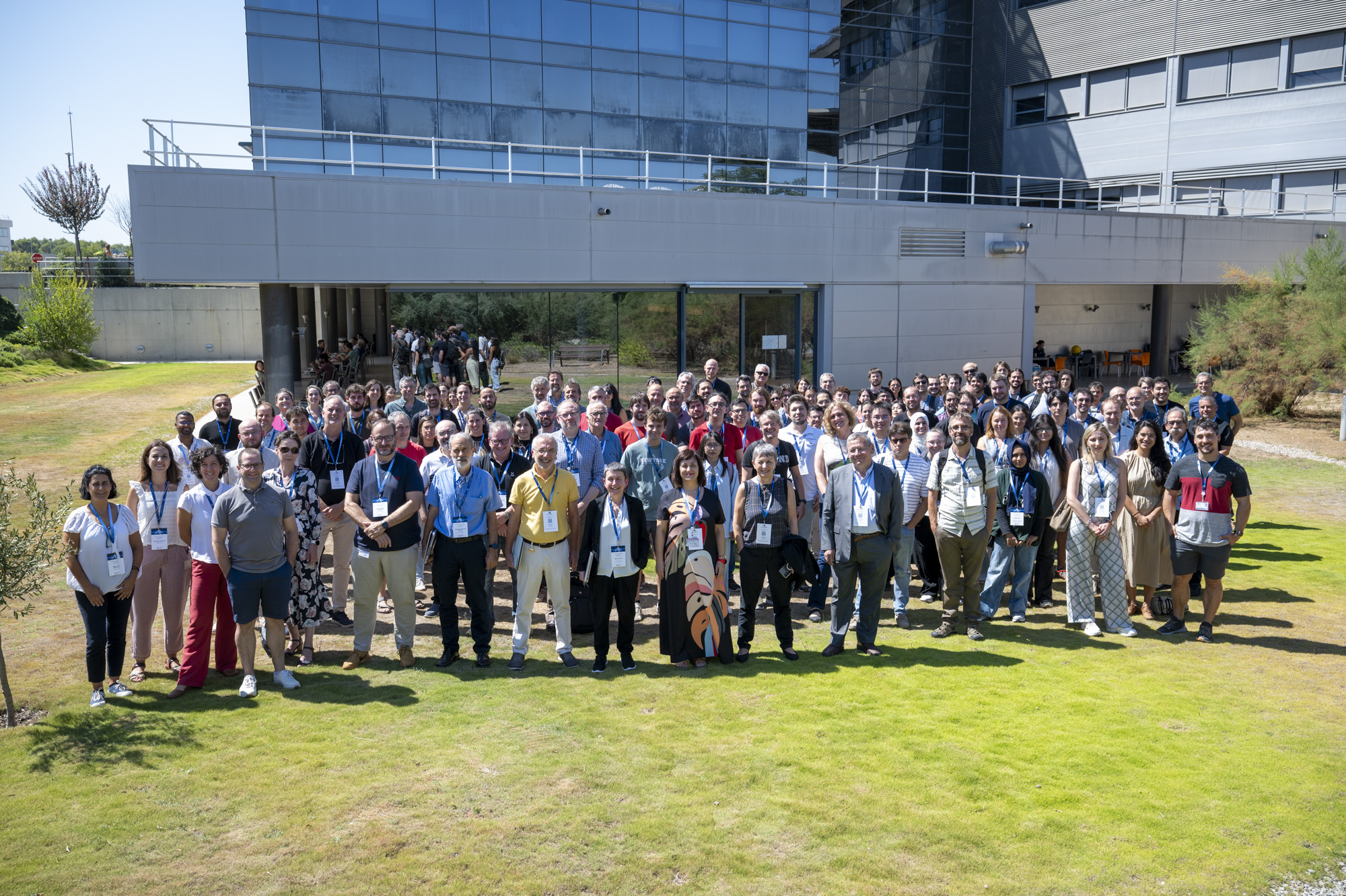
It has been a great pleasure to invite and welcome you to the ALBA Users' Meeting, which took place from 3rd to 4th of September 2026 at the ALBA Synchrotron in Cerdanyola del Vallès (Barcelona). It was a unique opportunity to learn more about ALBA’s new capabilities to support you in your research and provide a platform for communication and exchange within the community and with the facility.
Following the formal approval of the ALBA II project in 2025, this meeting has been an excellent occasion to share key updates on how ALBA II will transform the facility, including the beamline upgrades and new scientific capabilities.
The meeting included an overview of the ALBA II project, the science which will be enabled and the new tools that ALBA II will deliver to the community. An important aspect, the collaboration with the community to create new capabilities within the user program, was introduced and explained on the example of new instruments, available within the InCAEM facility, including a new TEM, a scanning microscopy and a High-Performance-Computing (HPC) platform with preconfigured workflows. The opportunities which come with InnoFAB, a new state-of-the-art Lab-in-FAB with 200mm wafer capabilities, currently constructed in the direct neighborhood of ALBA were also discussed. Early-career researchers play a vital role in shaping the future of our facility. In this context, we introduced a new Early Career Distinction Award to recognize outstanding scientific work conducted using ALBA Synchrotron instruments.
The second day we showed the impact of ALBA II, specifically the Beamline Upgrade Program, from the user’s perspective. These parallel sessions combined technical perspectives on the upgrades with insights into the enhanced scientific possibilities they will enable, featuring invited talks and contributions from leading experts across a range of disciplines.
This meeting has been developed in close collaboration with ALBA staff and the AUSE members, with the aim of delivering a dynamic and community-driven program.


ALBA Overview (From ALBA to ALBA II), Science & Partnerships, ALBA II Status
Based on the operational excellence, ALBA had gained over the past 15 years, the current upgrade of ALBA to a 4th generation light source is pushing the required sample size for spectroscopic and scattering techniques to the micrometer and sub-micrometer level and allows to strengthen imaging capabilities by optimizing existing instruments and adding coherent based techniques. The upgrade of the existing beamlines in combination with three new beamlines, to be operational after the one-year shutdown in 2032, is also adding high throughput and data analytics capabilities to the user program. Combined with enhanced operando and in-situ capabilities, ALBA is adding to the standard user program a high-resolution Transmission Electron Microscopy as well as a Scanning Surface Microscopy platform.
To give our users the full benefit of an optimized suite of instrumentation and integrating these tools into the Artificial Intelligence (AI) ecosystem, ALBA currently develops new ways of accessing these tools and collaborate with the facility to gain the methodologies you will need for your research or developments. A central part will be multimodal workflows for target areas, providing the user with a tool to reach the research goals in shorter time and in systems of increased complexity and inhomogeneity. Building on shared data spaces and data analytics, allowing not only the data provenance of data produced at ALBA, but correlating these with other data sets including proprietary data, will largely benefitting the connection between basic science and innovation; with an integrated IP generation- and access- management, workflows can be scaled and integrating various resources, promoting and optimal utilizing the Spanish and European innovation ecosystem.
Based on examples, the talk will explain the concept of workflows, demonstrating not only the power of this new tool but also showing the basic concepts of data and software management. It will also focus on the impact on existing and planned access modes as well as new ways to collaborate with the facility, independent if you are a researcher, a representant of a technology- or research-infrastructure, or an industrial player.
The upgrade of ALBA to a fourth-generation light source was approved in September 2025 and aims to ensure the competitiveness of the ALBA Synchrotron for the coming decades. The project includes the complete replacement of the storage ring, which will reduce the electron beam emittance from the current 4300 pm·rad to 200pm·rad. It also includes the upgrade of the existing beamlines to fully exploit the enhanced beam characteristics and the construction of new beamlines. Two of these new beamlines will be nanoprobe facilities, featuring source-to-sample distances exceeding 200m. This presentation will provide an overview of the current status of the various upgrade projects and their implementation schedule.
The In situ Correlative Facility for Advanced Energy Materials (InCAEM) is a new open-access research infrastructure that brings together complementary characterization techniques to study advanced energy materials under realistic operating conditions. Developed within the Spanish Complementary Plans Programme on Advanced Materials, InCAEM is a joint initiative of the ALBA Synchrotron, the Institute of Materials Science of Barcelona (ICMAB-CSIC), the Catalan Institute of Nanoscience and Nanotechnology (ICN2), and the Port d'Informació Científica (PIC-IFAE).
The facility combines state-of-the-art scanning transmission electron microscopy ((S)TEM), scanning probe microscopy (SPM), synchrotron-based techniques and dedicated sample environments to enable in situ and operando experiments across multiple length scales. By applying these complementary techniques to the same materials, InCAEM provides a correlative approach that links structural, chemical, electronic and functional information, offering a more comprehensive understanding of the mechanisms governing material performance.
In addition to its experimental capabilities, InCAEM integrates dedicated
computational resources for data management, multimodal data analysis and
workflow integration. This digital infrastructure complements the correlative experimental approach by enabling the efficient integration of datasets from multiple characterization techniques, supporting reproducible data processing and providing a unified framework for the analysis of complex in situ and operando experiments.
InCAEM is designed to serve both academic and industrial users working on
materials for batteries, electrocatalysis, photovoltaics and other energy-related technologies. By combining cutting-edge instrumentation with integrated data infrastructures and correlative methodologies, the facility aims to accelerate the discovery, optimization and validation of next-generation energy materials.
FaXToR is the first hard X‑ray micro‑tomography beamline operating at the ALBA Synchrotron, designed to deliver fast, high‑flux, multi‑scale imaging across a broad energy range. Its architecture combines filtered white and monochromatic beam modes, enabling absorption, propagation‑based phase contrast, and grating interferometry. A versatile endstation with multiple detection systems supports quasi‑simultaneous imaging at different spatial and temporal resolutions, advancing a multi‑scale, multimodal approach to investigate morpho‑structural changes across diverse materials [1]. A central focus of FaXToR is virtual histology, where phase‑contrast imaging and grating interferometry provide soft‑tissue sensitivity comparable to classical histology but without sectioning or staining. Sub‑micrometre voxel sizes, long propagation distances, and high‑flux illumination enable visualization of cellular‑scale features within intact organs, supporting quantitative 3D morphometry and pathology‑driven analysis. This workflow is strengthened by the beamline capability for dynamic studies, capturing physiological or mechanical processes in real time. The optical layout is centered on a double multilayer monochromator optimized for vibrational stability and high‑power handling. Advanced filtering units, diagnostics, and fast shutter systems ensure precise dose control and high‑speed acquisition. The experimental hutch hosts a flexible tomography tower accommodating diverse in situ environments, while dual microscope detection platforms provide wide‑range spatial resolution. High‑performance computing pipelines enable on‑the‑fly reconstruction and real‑time visualization. Commissioning activities validated performance in terms of power, flux, spectral stability, and monochromator calibration. Applications span biomedicine, paleontology, materials science, geoscience, and energy storage. FaXToR establishes a strategic foundation for future multiscale imaging developments at ALBA and aligns with the long‑term upgrade path toward ALBA II.
The BL06-XAIRA beamline is the microfocus macromolecular crystallography (MX) instrument at the ALBA synchrotron, that just accomplished its first year of User Operation.
XAIRA currently delivers a highly stable, intense X-ray beam of 4 × 3 $µm^2$ (FWHM at 1 Å wavelength) in the energy range of 4.0 to 14 keV, specifically tailored for demanding structural biology projects. This high-flux beam is paired with a state-of-the-art Dectris Eiger2 XE 9M photon-counting detector capable of acquiring data at frame rates up to 1 kHz. This configuration enables rapid data collection and supports time-resolved serial synchrotron crystallography (SSX) experiments within the millisecond regime.
Another differential trait of XAIRA is its specialized end-station chamber, in which the entire experimental setup —including the sample environment, detector, and cryostream— is enclosed within a controlled helium atmosphere. Operating in helium prevents flux attenuation at low energies, improves the signal-to-noise ratio of diffraction images at all photon energies, and mitigates the vibrations induced by the cryogenic stream blowing the crystal. Preliminary results with lysozyme show that, in a “worst case scenario” of a big crystal (100 µm per side), photon energy of 12,661 keV and short (76mm) sample-detector distance, at 1 Å resolution the signal-to-noise ratio increases by a factor of 3 and the resolution improves by ~0.1 Å. The chamber encasing the end stationgrants full compatibility with standard MX sample formats and conventional operation in air, while the associated circuit allows for full recovery of helium.
Since welcoming its first official users in July 2025 and its transition into full operation, XAIRA has demonstrated its value for challenging structural biology projects. Here we present case examples of successful results, describe how the beamline can be used to obtain the best data set for different types of challenging samples, and how the automated data processing software is used to provide immediate feedback and guide the data collection decision-making.
We will also discuss the future unique experiments enabled by the routine and automated operation of XAIRA in helium, and present the foreseen upgrades to be implemented in the upcoming ALBA-II Beamlines Upgrade Plan, which will improve both the beamline optics system and the End Station capabilities.
Only a limited number of experimental techniques can provide detailed information about interfaces under relevant working conditions, including controlled pressure, temperature, electrical potential, illumination, in the presence of gas or liquid environments. Among these, photoelectron spectroscopy, surface X-ray diffraction, and X-ray scattering techniques are particularly valuable for investigating complex interfacial systems. They provide complementary information on the chemical, electronic, and structural properties of the outermost atomic layers while enabling, in many cases, measurements under operando conditions.
In this talk, we will present the current and near-future capabilities available at the ALBA Synchrotron for performing these types of experiments, with particular emphasis on the experimental setups that enable the study of working interfaces under relevant environmental and electrochemical conditions. To illustrate these capabilities, we will discuss selected examples from different research areas, demonstrating how synchrotron-based techniques can provide unique insights into the structure, composition, and electronic properties of interfaces during operation. We will also highlight the advantages of combining complementary X-ray and photoelectron spectroscopy approaches to establish structure–property relationships and unravel the dynamic processes governing complex interfaces.
Angle-resolved photoemission spectroscopy (ARPES) is the premier technique for determining the electronic band structure of solids and has found wide application across many classes of materials, including oxides, semiconductors, metals, and low-dimensional materials and surfaces. Among the important topics it addresses are the many-body interactions that underpin the ground- and excited-state functionalities of materials. Recently, the development of nanoARPES using microfocused X-ray beams has opened ARPES to a wider class of samples and enabled in situ measurements of 2D devices under applied electric fields, currents, and strain, as well as modification with focused femtosecond laser pulses. In this talk I will give an introduction to the ARPES technique and the MAESTRO facility, and share some of our recent work on the band structure and many-body interactions in 2D heterostructures of chalcogenides, graphene, and boron nitride, and on light-induced metastable phases in 1T-TaS₂.
Gels can be defined as composite materials resulting from the combination of a solid (gelator) capable of generating a three-dimensional network that retains the flow of a much larger quantity of fluid due to surface tension. In particular “supramolecular gels” are an increasingly important class of new materials, defined by the ability of the molecular components to self-assemble through non-covalent or supramolecular interactions, generating a three-dimensional network that yields to gels. Importantly, these gels can be tailored to present optical and chiroptical activities,[1] to obtain composite or hybrid materials,[2,3] and to be biocompatible or have therapeutic activity.[4] Due to the fact that the nature of the interactions maintaining the network is dynamic and non-covalent the gelation is reversible in most of the cases. Hence, one of the most pursued characteristics when rationally designing these systems is the responsiveness to external factors such as temperature, mechanical stress, light, chemical agents, enzymes, and pH.[5] Of all of these, the use of light to induce transformations in gels is particularly interesting because it is non-invasive, can be remotely applied, and, if the alteration is reversible, does not change the chemical composition. But, when rationally designing a supramolecular building block for these new materials, it is crucial to rationally design the self-assembly interactions to foster gelation over crystallization because there is a delicate balance for a low-weight molecule to behave as a gelator or as a crystal building block. A balance that could be tuned by changing the experimental conditions during system decomposition and phase formation.[6]
We propose a dual‑beamline, multimodal approach using XALOC and NCD‑SWEET on the same samples to study self‑assembly across multiple length scales, with the aim of elucidating the physicochemical mechanisms that govern this balance. For that, we studied a model system capable of developing reversible trans-to-cis and cis-to-trans transitions upon irradiation with light sources of different energy (365 nm and 520 nm, respectively). Remarkably, as this molecule developed crystalline gels under specific conditions, this photoresponsive feature allowed for the study of simultaneous single-crystal-to-liquid crystal and gel-to-sol transitions, and vice versa. To have a clear insight into the structure-property relationship, we performed high quality single crystal X-ray diffraction studies, which successfully led to the determination of the molecular structure of the systems of interest. Even more, we also conducted SAXS and WAXS experiments to depict a clearer panorama of all the structural changes that are taking place while irradiating with different light sources. The necessity of performing fast data-acquisition during short periods of time to correctly follow the self-assembly process while introducing external irradiation sources into the sample cabinet to account for the stimuli-responses studies made the use of cutting-edge facilities as the BL11 - NCD-SWEET and BL13 - XALOC beamlines mandatory. We believe that this example reflects the synergic nature of these two beamlines working together to tackle novel challenges in crystallography and new materials.
Following cardiac injury, mammals are unable to regenerate the affected tissue and instead form a fibrotic scar to preserve mechanical function. In contrast, zebrafish possess the remarkable capacity to fully regenerate the damaged area within 30 days post-injury.
The extracellular matrix (ECM) plays an essential role in this regenerative process. Using high-resolution mechanical measurements (atomic force microscopy and nanoindentation), our group identified a gradient in ECM stiffness across the regenerating tissue. This stiffness gradient arises from variations in the crosslinking of collagen fibers in a process mediated by periostin b. These findings indicate that the mechanical properties of the ECM are dynamically regulated during zebrafish heart regeneration, potentially playing a critical role in guiding cellular behavior and tissue remodeling.
To better understand whole-heart structure and morphology without prior physical manipulation (such as decellularization or sectioning, which can disrupt native architecture), we applied synchrotron-based X-ray Phase Contrast Imaging (X-PCI) at the FAXTOR beamline of the ALBA Synchrotron facility. This technique uses highly intense X-rays to visualize soft tissues in 3D at microscopic resolution without the need for contrast agents or destructive preparation methods. We examined cardiac morphology, ECM organization, and structural differences between wild-type zebrafish (which regenerate normally) and postnb KO mutant zebrafish (which lack the postnb gene and are unable to regenerate their hearts). We analyzed adult zebrafish hearts at 7 days post ventricular amputation or sham operation (non-amputated controls), preparing formalin-fixed, paraffin-embedded (FFPE) heart blocks for imaging. Image acquisition was performed at 20 keV with a voxel size of 0.65 µm.
In our first beamtime session, we acquired high-resolution tomographic images from 9 zebrafish hearts, including WT and PostnB KO mutant animals from both non-amputated hearts and hearts at 7 dpa. Despite the modest number of successfully acquired samples, we obtained valuable preliminary results. We observed that non-amputated wildtype hearts exhibit more structured cardiomyocyte fibers/trabeculae, a thicker epicardium/cortical cardiomyocyte layer, and appear more organized overall compared to non-amputated PostnB mutant hearts. Furthermore, analysis of one heart per amputated condition showed that, at 7 dpa, the wildtype regenerating region was organized and of similar dimensions to the original myocardium, whereas the PostnB mutant heart formed a disorganized, blob-like mass and larger than the original tissue (Figure 1). Further studies are needed to fully characterize the native organization of the myocardium, as well as the precise dynamics of its regrowth.
3Sbar (Surface Structure and Spectroscopy at bar pressure) at ALBA Synchrotron is the new X-Ray micro-spot beamline for studying surfaces and interfaces under realistic operating conditions. By integrating NAPHAXPES,XAS, and advanced scattering techniques (SXRD, GIXD, and GISAXS), it enables simultaneous chemical and structural characterization in gases or liquids. Operating between 3 and 17 keV and from ultra-high vacuum to several bars of partial pressure at the sample.
INTRODUCTION
Structural deformations in zeolites during adsorption processes have been previously reported for materials such as MFI [1], RHO [2], AlPO-LTA [3], or AlPO-CHA [4]. These changes are typically associated with an increase in adsorption capacity above a certain pressure, as evidenced by the presence of steep steps in the adsorption isotherm. Such behavior is often linked to second-order phase transitions, where atomic connectivity is preserved, but changes in symmetry may occur. These transitions depend on both the pressure and the nature of the adsorbate.
Here, we describe the structural response of pure silica zeolite ZSM-11, also known as silicalite-2 [5] (IZA code: MEL), during the adsorption of light hydrocarbons (propane and butane). This study combines in situ powder X-ray diffraction (PXRD) with adsorption isotherm measurements.
RESULTS AND DISCUSSION
Propane and butane adsorption isotherms at 298 K do not present abrupt steps (Figure 1, left), in contrast to Ar adsorption at 77-97 K, which exhibits a sharp step, as previously reported (Figure 1, right) [6]. This suggested the absence of phase transitions during hydrocarbon adsorption in silicalite-2 and a continuous deformation of the framework.
Figure 1: (Left) hydrocarbon adsorption isotherms at 298 K; red: propane, black: butane; (right) Ar adsorption isotherms at 77 K (black), 87 K (red), 92 K (green) and 97 K (blue) on silicalite-2
To further analyze these phenomena, in situ PXRD experiments were conducted under atmospheres with controlled partial pressures of the selected hydrocarbons diluted in helium at room temperature.
All diffraction patterns of silicalite-2 obtained at different partial pressures were indexed with the same tetragonal symmetry (I -4 m 2), indicating that there were no symmetry changes during the filling of the microporous space of the zeolite. However, a continuous increase in the unit cell parameters was observed when increasing the ratio of hydrocarbon in the gas flow (Figure 2). This expansion is not linear with respect to the hydrocarbon uptake, showing a small variation of the cell volume after the inclusion of the first molecules, which occupy the void spaces, that becomes larger once the pores are almost full (Figure 3). These results demonstrate that silicalite-2 exhibits a significant framework flexibility, accommodating hydrocarbon molecules through continuous expansion of the pore system while preserving its structural symmetry. Moreover, this process is fully reversible, returning to the original cell parameters when submitting the material to a flux of pure helium at room temperature.
Figure 2: Variation of the unit cell parameters of silicalite-2 with the hydrocarbon/helium ratio (XCn). Red: propane; black: butane
Figure 3: Expansion of the cell volume of silicalite-2 with the uptake of hydrocarbon molecules. Red: propane; black: butane
EXPERIMENTAL
Silicalite-2 was synthesized following a previously reported procedure [7]. Prior to adsorption measurements the calcined material was degassed at 400 ºC under high vacuum for 12 h. Ar adsorption isotherms were measured using an ASAP-2020 (Micromeritics) equipped with a Cryotune system (3P Analytics), and hydrocarbon adsorption isotherms were measured using a BELSORP Max-II instrument. PXRD experiments were performed in an Anton-Paar XRK-900 reaction chamber attached to an Empyrean X-ray diffractometer. Prior to the measurements, the calcined material was degassed at 400 ºC under a continuous flow of helium. Hydrocarbon/helium mixtures with different proportions were then fluxed through the sample. Diffraction patterns were collected after equilibrium was reached for each flux composition.
CONCLUSIONS
Although zeolite flexibility is often associated with phase transitions driven by cation movement or structural/symmetry rearrangements, the present study shows that silicalite-2, which possesses no cations on the channel system, responds differently to light hydrocarbon adsorption. No phase transitions were observed. Instead, the material undergoes a continuous elastic expansion of the unit cell. This behavior highlights the intrinsic flexibility of the MEL framework, which adapts to guest molecules through smooth structural deformation while preserving its symmetry. Further studies using synchrotron radiation are expected to improve the structural resolution and gain a deeper insight into the adsorption-induced flexibility and the atomic displacements in the framework.
REFERENCES
[1] E. García-Pérez, J.B. Parra, C.O. Ania, D. Dubbeldam, T.J.H. Vlugt, J.M. Castillo, P.J. Merkling and S. Calero (2008) J. Phys. Chem. C, 112, 9976–9979.
[2] M. Palomino, A. Corma, J.L. Jordá, F. Rey and S. Valencia (2012) Chem. Commun., 48, 215–217.
[3] A. Krajnc, J. Varlec, M. Mazaj, A. Ristic, N. Zabukovec Logar and G. Mali (2017) Adv. Energy Mater., 7, 1601815.
[4] J. Varlec, A. Krajnc, M. Mazaj, A. Ristic, K. Vanatalu, A. Oss, A. Samoson, V. Kaucic and G. Mali (2016) New J. Chem., 40, 4178–4186.
[5] D. Bibby, N. Milestone, L. Aldridge (1979) Nature 280, 664–665
[6] V. Sánchez-Gil, E. G. Noya, J. M. Guil, E. Lomba, S. Valencia, I. da Silva, L. Pusztai, L. Temleitner (2016, J. Phys. Chem. C, 120, 4, 2260–2270
[7] O. Terasaki, T. Ohsuna, H. Sakuma, D. Watanabe, Y. Nakagawa and R.C. Medrud (1996) Chem. Mater., 8, 463–468.
ACKNOWLEDGEMENTS
The authors thank the financial support of the Spanish Ministry of Science and Innovation (CEX2021–001230-S and PID2022-136934OB-100 grants funded by MCIN/AEI/10.13039/ 501100011033 funded by “ERDF A way of making Europe” and TED2021-130191B-C41 grant funded by the European UnionNext Generation EU/PRTR) and the Generalitat Valenciana (Prometeo 2021/077 and CIPROM/2024/050).
BL16 NOTOS bridges the gap between X-ray Absorption Spectroscopy (XAS) and X-ray Diffraction (XRD) by enabling sequential, quasi-simultaneous measurements on a single sample. This multimodal approach provides a comprehensive picture of material evolution: operando XAS offers element-specific sensitivity to monitor oxidation states and local coordination changes, while operando XRD provides direct evidence of long-range structural transformations, phase transitions, and lattice strains. Correlating these electronic and structural dynamics is vital for elucidating fundamental structure-function relationships and failure mechanisms across diverse scientific disciplines. This is particularly crucial in catalysis and energy materials research, where understanding complex electrochemical processes during operation is essential for improving performance and lifespan.
To facilitate these advanced studies, NOTOS features an extensive sample environment pool equipped with specialized multi-cell battery supports, high-temperature catalysis reactors, and real-time gas and liquid analysis systems.
Further enhancing its technical capabilities, the commissioning of the Kirkpatrick-Baez (KB) mirror system was successfully concluded in July 2026. This new micro-focus station delivers a stable, micro-focused beam, reaching spot sizes as small as 10 x 10 μm² (VxH) at the sample position. In the InCAEM (In Situ Correlative Facility for Advanced Energy Materials) framework, this instrument will open significant possibilities for correlative in-situ and operando experiments: researchers will study the identical sample area across NOTOS and the Scanning Transmission Electron Microscope in JEMCA, obtaining complementary information at multiple length scales without sample alteration.
Combining two active materials in a single electrode is an established strategy to improve lithium-ion battery performance, reduce cost, and tailor properties such as the voltage profile and thermal stability [1,2,3]. Many commercial EVs employ blended positive electrodes (e.g., NMC–LMO, where NMC is LiNiₓMnᵧCozO₂, x+y+z = 1, and LMO is LiMn₂O₄) and negative electrodes (e.g., silicon–graphite). However, the origin of the synergistic behavior of blends remains poorly understood, and their design is largely empirical. Furthermore, very few studies address the effect of temperature on such systems, a critical factor, since EV batteries must operate over a wide and constantly varying temperature range.
Here we investigate the temperature dependence of the reaction mechanism in NMC–LMO blended cathodes, one of the most commercially relevant blend chemistries. Operando synchrotron X-ray diffraction at the MSPD beamline and operando X-ray absorption spectroscopy at the CLAESS beamline were performed on 3 coin cells at 0ºC, 25ºC and 45ºC. XRD follows the structural evolution of each phase individually as seen in Figure 1 for NMC (003) and LMO (111) reflections. In the XANES data the Mn K-edge was analyzed by multivariate curve resolution (MCR) the redox response of which is dominated by LMO. Both techniques reveal a clear alteration of the reaction mechanism with temperature. At elevated temperature, NMC shows a more solid-solution-like behavior on both charge and discharge, as well as a larger c-axis contraction at the end of charge, which could impact its structural stability upon extended cycling. At 0 °C, the LMO (111) reflection develops a markedly broader distribution of lattice parameters, indicating a more inhomogeneous reaction across the electrode (Figure 1). Most strikingly, both XRD and the MCR-resolved XANES components (Figure 2) show that at 0 °C, during the first charge, it is LMO that reacts first, despite operating at higher potentials and therefore being expected to react at a later stage. To the best of our knowledge, this temperature-induced inversion of the reaction sequence has not been previously reported.
These results provide mechanistic insight into how blended electrodes function away from ambient conditions, contributing to both the fundamental understanding of such synergistic systems as well as testing the systems in conditions closer to their real applications. In addition, this study serves as a proof of concept for the simultaneous measurement of three coin cells at three different temperatures, with a setup [4] adapted to both MSPD and CLAESS, increasing the throughput and ease of temperature-dependent operando studies.
Figure 1: Operando X-ray diffraction patterns showing the (003) reflection of NMC (lower angles) and the (111) reflection of LMO (higher angles). A clear difference in the reaction mechanism is observed as temperature is varied.
Figure 2: Left: MCR-ALS spectral components resolved from the operando Mn K-edge XANES. Middle and right: concentration profiles of the two components during cycling of NMC–LMO cells at 0 °C, 25 °C, and 45 °C, together with a pure LMO reference.
References
[1] J. Smith et al 2012 J. Electrochem. Soc. 159 A1696
[2] Hideki Kitao et al 2005 Electrochem. Solid-State Lett. 8 A87
[3] ACS Appl. Mater. Interfaces 2016, 8, 31, 20147–20156
[4] R. G. Houdeville et al 2024 J. Electrochem. Soc. 171 020533
Within the ALBA Life Sciences Section, current efforts focus on establishing a multi-scale, multi-technique imaging strategy to map biological systems across hierarchical levels—from cells to tissues, and from tissues to whole organisms. The Fast X-ray Tomography and Radioscopy beamline (FaXToR) is central to this vision, offering a dedicated hard X-ray µCT instrument for morphological characterization at the micrometer and sub-micrometer scales within large biological volumes. Operating at 8–70 keV with high photon flux and flexible detection configurations, FaXToR supports both static and time-resolved imaging to track dynamic physiological processes, biomaterial interactions, and pathological micro-architecture. In addition to propagation-based phase-contrast imaging, the beamline incorporates differential phase and dark-field imaging via Talbot grating interferometry. These modalities significantly enhance sensitivity in weakly absorbing soft tissues, enabling the non-destructive visualization of micro- and nano-structural features crucial for preclinical and fundamental medical research. By utilizing the beamline's multi-scale detection system, researchers can seamlessly bridge organ-level morphology with sub-micron structural detail. This presentation will highlight FaXToR’s emerging bioimaging capabilities, correlating them with the broader multimodal X-ray approaches in the ALBA portfolio. Furthermore, we will demonstrate how combining complementary phase-contrast modalities provides richer insights into tissue organization and function. Looking ahead, planned upgrades toward a 4th-generation synchrotron and expanded phase-contrast techniques will further strengthen ALBA's life-science imaging pipeline, unlocking unprecedented opportunities for dynamic, high-sensitivity biological characterization.
Aqueous Zn–MnO₂ batteries involve complex reaction processes during operation, including deposition/dissolution of the MnO₂-based electrode and zinc-containing species, and possible generation of side products. Understanding these material transformations under working conditions is essential for clarifying the reaction mechanism and degradation pathways of this battery system.
In this work, operando synchrotron X-ray diffraction was used to investigate the cathode evolution of aqueous Zn–MnO₂ batteries during electrochemical cycling. The XRD data provides direct structural information on the appearance, disappearance, and evolution of crystalline phases during battery operation. However, their interpretation is challenging due to the highly defective nature of MnO₂, the strongly crystalline zinc-containing by-products, and background contributions from the operando cell. Intense reflections from zinc hydroxysulfate-related species can mask weaker diffraction changes associated with the MnO₂ electrode and other evolving phases.
To assist the analysis, Multivariate Curve Resolution was used as a data-preprocessing tool rather than as a direct phase-identification method. MCR was applied to separate the dominant zinc hydroxysulfate-related diffraction contribution, which was then subtracted from the original patterns according to its evolution profile, followed by intensity renormalization to reduce background distortions. This treatment improves the visibility of weaker and broader diffraction peaks. The highlighted changes indicate relevant composition evolution of the Mn oxide during electrochemical cycling. This effect overlaps with the expected deposition-dissolution mechanism, which would simply show changes in intensity. These experiments expand our understanding of the dynamic material evolution in aqueous Zn–MnO₂ batteries.
Go through the attached file
Since 2013 CLAESS offered access to x-ray absorption and x-ray emission spectroscopy to a wide user community, running in a regime of around 50 experiments per year, most of them addressing key social challenges. The main focus on catalysis, energy related materials, environmental science, and solid-state physics resulted in the development of dedicated setups, optimized experimental strategies, and staff member scientific experience in the respective fields.
The gained experience, together with some users’ necessities allow to identify, in the incoming ALBA II upgrade, interesting opportunities. Indeed, in the framework of the machine upgrade an upgraded CLAESS beamline could significantly expand its capabilities by providing nearly simultaneous access to μXRF, μXAS, and μXES with micrometer spatial resolution and ~1 s full-scan acquisition times.
The proposed upgrade includes new optics, a faster monochromator and sample stage, together with enhanced standard fluorescence detection and highly efficient X-ray emission spectrometers. These improvements will enable high-throughput operando and in-situ experiments with 100-fold better time resolution while minimizing radiation damage. Moreover, both environmental science and solid-state physics will profit of the higher space resolution allowing experiments which are currently requesting access to other facilities. Finally, enhanced detection limits will further extend applications to diluted systems.
The upgraded CLAESS beamline is expected to strongly support ALBA II’s key pillars — multi-modal and multi-scale programs, high-throughput techniques, and operando/in-situ studies — with direct impact on research in renewable energy, green catalysis, batteries, environmental science, and food security, and will enhance ALBA’s competitiveness for studies of heterogeneous and dynamic systems under realistic working conditions.
Bacterial quorum sensing (QS) is a cell-to-cell communication with a central role in the virulence of several high-priority pathogenic bacteria, such as Pseudomonas aeruginosa or Acinetobacter baumannii. Degradation of the mediator signal, N-acyl-L-homoserine lactones (AHLs) in Gram-negative bacteria, is an attractive approach to develop alternative antibacterial treatments. Moreover, different pathogens produce different variants of AHLs, which vary on the acyl chain length and/or oxidation state of the C3, which could potentially open the possibility for tailored interventions.
Several natural lactonases can hydrolyze AHLs, although they are usually low-specificity and/or do not possess appropriate biophysical properties such as thermostability, proteolytic resistance or pH tolerance. Even though enzymes possess an enormous structural flexibility that permits to adapt them to new functions and properties, this same flexibility and complexity make their rational redesign highly challenging, even with the recent and continuous advances in experimental and -especially- computational approaches
To overcome these challenges, we developed a computational-experimental framework for enzyme design and optimization integrating biochemical validation experiments into a fully customizable, computational pipeline to generate an iterative refinement process. Starting from AidH, a structurally-known broad-spectrum, non-thermostable lactonase, we computationally generated 15 optimized designs that combine backbone redesign (RFDiffusion3) of the substrate entry pocket with an iteratively optimized sequence (LigandMPNN, AlphaFold3, Rosetta, and PLACER) for the redesigned region biasing substrate preference toward short-chain AHLs, as well as evaluating the substrate stability in the active site using molecular dynamics simulations. Experimentally, we established a bioassay to evaluate the activity of these enzymes and validate it with the wild-type AidH and the catalytically inactive variant AidHS102G. We now aim to biochemically characterize and structurally resolve these variants to obtain a deeper understanding on the structural determinants of substrate specificity to allow rational, substrate-specific redesign of lactonases targeting the AHL profiles of priority pathogens.
The ultrastructure of biological tissues holds cues to critical answers impacting society across multiple domains – from neuroscience to cancer, agriculture or host-pathogen interactions in infectious diseases. Moreover, combining readouts of tissue ultrastructure with functionally relevant insights can be used to accurately interrogate how multicellular systems operate – such as how specific neurons in a neuronal circuit compute information [1, 2]. However, efficient platforms to gather this empirical insight are missing, leaving a large fraction of this research potential untapped. Volume electron microscopy [3, 4] requires slicing the sample sequentially every tens of nanometres making it challenging to address volumes equal or larger than a cubic millimetre [5, 6]. In turn, coherent X-ray imaging can resolve tissue ultrastructure without the need of slicing [7], and 4th-generation synchrotrons provide sufficient flux to make mm3 nanoscale imaging a routine approach available to any research laboratory worldwide [8]. The ALBA synchrotron has started the construction of CORUS, a dual-branch beamline providing capacity for tissue nanoimaging, fluorescence and cryospectroscopy. It will use beams of 10, 19 and 27 keV, and operate under both room temperature and cryo regimes. The nanoImag endstation will be designed for X-ray holographic nanotomography [9, 10] of up to 1 mm3 volumes at ultrastructural resolution, and 3D X-ray fluorescence. The nanoFluoSpec endstation will resolve elemental compositon with subcellular resolution using 3D X-ray Fluorescence and it will discriminate chemical species using X-ray spectroscopy at two energy ranges (4-10 keV and 10-25 keV). Together, CORUS will offer nanoimaging, elemental mapping and speciation with a built-in design for correlative multimodal and multiscale explorations of the structure and function of cells and tissues. It will be useful for multiple areas of academical and industrial research, such as the study of metallomics, pathogen-host interactions, metal nanocomposites, toxicology, biomineralization, bioremediation strategies, food security and sustainable agriculture, and nanoimaging of biological tissues across the plant and animal domain including human clinical biopsies, and revealing connectomes when addressing neuronal tissues.
References
[1] - K. L. Briggman, M. Helmstaedter, and W. Denk, Nature, 471(7337), 183-8 (2011).
[2] - J. A. Bae, M. Baptiste, M. R. Baptiste et al., Nature, 640(8058), 435-447 (2025).
[3] - W. Denk, and H. Horstmann, PLoS Biol, 2(11), e329 (2004).
[4] - C. J. Peddie, C. Genoud, A. Kreshuk et al., Nature Reviews Methods Primers, 2(1), 51 (2022).
[5] - L. F. Abbott, D. D. Bock, E. M. Callaway et al., Cell, 182(6), 1372-1376 (2020).
[6] - G. S. Jefferis, L. Collinson, C. Bosch et al., Wellcome Trust, (2023).
[7] - C. Bosch, T. Aidukas, M. Holler et al Nature Methods, (2025).
[8] - M. Du, Z. W. Di, D. Gursoy et al., J Appl Crystallogr, 54(Pt 2), 386-401 (2021).
[9] - A. T. Kuan, J. S. Phelps, L. A. Thomas et al., Nat Neurosci, 23(12), 1637-1643 (2020).
[10] - Y. Zhang, C. Bosch, T. Ackels et al., bioRxiv, 2025.04.24.650439 (2025).
Cell division is driven by the mitotic spindle, a self-organized macromolecular machine that assembles in prophase. During metaphase, it comprises microtubule (MT) kinetochore fibers connecting chromosomes to the bipolar MT network of the spindle, and astral MTs extending from the poles to the cell cortex. With the onset of anaphase, the mitotic spindle undergoes significant rearrangement. Shortening of kinetochore fibers moves the sister chromatids toward the poles, separating the chromosomes. Simultaneously, several motor and MT-associated proteins relocate to interpolar MTs, bundling their antiparallel plus ends at the central spindle (or midzone), essential to maintain proper spindle architecture and ensure accurate chromosome segregation. In vitro studies using purified proteins have identified the central spindle proteins PRC1 and KIF4A as a minimal set of components capable of organising MTs into bundles with central antiparallel MT plus-end overlaps that resemble anaphase midzone bundles. Their organisation was previously visualised using fluorescence microscopy. To gain higher-resolution insights into their three-dimensional architecture, we modified the procedure of minimal midzone bundle self-organisation to allow their visualisation by cryo-electron tomography (cryo-ET). We reduced the size of the reconstituted minimal midzone bundles while preserving other characteristic midzone features. Preliminary data indicate that we can now successfully image these reconstituted anaphase-like bundles using cryo-ET. This approach will enable a more detailed analysis of microtubule arrangements in reconstituted dense microtubule networks moving forward.
De novo protein enables the creation of novel structures of stable protein structures that exceed those found in nature. However, designing antibody-like scaffolds that combine stability with functional versatility remains a major challenge, constrained by the conserved β-sandwich architecture of immunoglobulin (Ig) domains and designing functional loop regions.
Our group has developed hyperstable, non-natural Ig-domain dimers, validated by X-ray crystallography, that act as robust frameworks for functional loop insertion. Building on these validated scaffolds, we are exploring the possibility of engineering two-domain Ig scaffolds to contain functional cavities at the interface between domains with antigen recognition loops. We hypothesise that the controlled incorporation of small-molecule binding pockets and antigen-binding loops will generate stable and controllable frameworks for performing programmable binding functions.
To address this issue, we are implementing a multi-step computational pipeline for designing a library of Ig-domain pairs with engineered inter-domain pockets and exploring the de novo antigen-binding loop design in those validated two-domain Ig frameworks. AI-based and physic-based tools are used for backbone - sequence design, structure prediction, modelling and interface optimization, followed by in silico evaluation. The most promising candidates will be recombinantly expressed in E. coli and experimentally characterized using SEC-MALS, circular dichroism, SPR/BLI, and X-ray crystallography to validate their structure and function. This will establish a versatile platform for future immunoglobulin-like scaffolds customized to bind protein targets of medical interest.
The high photon flux and energy tunability of synchrotron facilities provide a clear advantage for the characterization of Atomic Layer Deposition (ALD) processes. A mobile custom-built ALD reactor (ALD2) has been developed at the ALBA Synchrotron to enable in situ monitoring of film growth using, up to now, two X-ray-based techniques: Grazing Incidence Small Angle X-ray Scattering (GISAXS) at NCD-SWEET (BL11) and X-ray Absorption Spectroscopy (XAS) at NOTOS (BL16).
The setup consists of a high-vacuum, pump-type reactor compatible with multiple precursors and oxidants, therefore enabling the combination of different ALD coatings during the same experiment. It achieves a base pressure of 10–6 mbar (limited by the semi-transparent windows used for the X-ray measurements, i.e. Kapton and mica) and can also operate under rough vacuum (10–3-10–2 mbar). The mobile reactor is a modular system that can be accommodated for different specific geometries: for GISAXS, two CF40 flanges with 50 µm-thick mica windows allow the passage of incident, reflected and scattered X-rays along the beam direction, being recorded with the SAXS detector (Pilatus3 S 1M) placed at 6.5 m; while for XAS, the two CF40 are rearranged in a perpendicular orientation in the horizontal plane and changed by 25 µm-thick Kapton windows, enabling measurements covering all the beamline energy range (4.7 - 30 keV), with the fluorescence detector used (Silicon Drift Detector with 13 channels from Canberra Olen) which is located at 90° with respect to the X-ray beam (and the sample at around 30° with respect to the incident beam).
The reactor was validated with in situ deposition of TiO2 thin films using the TTIP/H2O at 100, 150 and 200 °C as a deliberately demanding test case given its low growth per cycle (0.17 Å·cycle⁻¹). At NCD-SWEET, real-time GISAXS monitoring over 1000 ALD cycles resolved the progressive evolution of film morphology through model-independent analysis of the diffuse scattering signal, capturing the full sequence from the first cycles to steady-state growth. At NOTOS, in situ Ti K-edge XANES analysis of the edge jump evolution quantitatively identified three kinetically distinct growth regimes, with linearity in the steady-state regime confirming the self-limiting character of the process. The combination of both techniques provides a detailed picture of TiO2 ALD growth dynamics: with XAS capturing early chemical nucleation and GISAXS tracking morphological evolution, demonstrating the potential of the ALD2 reactor for multi-technique in situ characterisation of ALD processes at ALBA Synchrotron.
Trigonal PtBi₂ has emerged as a compelling platform for studying the interplay between crystal symmetry, topology, and superconductivity [1,2,3]. In this noncentrosymmetric Weyl semimetal, the absence of inversion symmetry and strong spin–orbit coupling give rise to topological surface states, while superconducting signatures appear to be strongly inhomogeneous from the nano- to the microscale and, remarkably, confined to the surface Fermi arcs [4,5]. This unusual behavior suggests that the superconducting properties of PtBi₂ may be intimately linked not only to its electronic topology, but also to subtle structural distortions and local symmetry breaking within its layered lattice. Understanding how these local structural degrees of freedom evolve is therefore essential for clarifying the origin of its unconventional superconducting response.
This complex scenario requires a combined approach capable of disentangling the structural and electronic ingredients that contribute to the unique behavior of PtBi₂. In this work, we use temperature-dependent X-ray absorption spectroscopy (XAS) and submicron spatially resolved angle-resolved photoemission spectroscopy (nanoARPES) to correlate local structural distortions with the surface electronic properties. The XAS results reveal an anomalous temperature evolution of the local atomic environment, characterized by two distinct upturns around 200 K and 60 K, possibly indicating the onset of competing or intertwined ordering tendencies that shape the low-temperature properties of PtBi₂. Notably, recent scanning tunneling spectroscopy and second harmonic generation studies have suggested that the anomaly near 60 K may be connected to real onset of surface superconductivity, whose spectroscopic signatures are otherwise detected at much lower temperatures [6,7]. Complementary nanoARPES measurements further reveal spatial fluctuations of the surface electronic states that correlate with local variations of the Bi-terminated surface, with a distinct change in the electronic response across the 60 K anomaly. These results point to a close connection between local lattice distortions, surface electronic inhomogeneity, and the unconventional superconducting phenomenology of trigonal PtBi₂.
References:
[1] K Takaki et al. (2022). Journal of the Physical Society of Japan, 91(3), 034703.
[2] A. Kuibarov et al. (2024). Nature, 626(7998), 294-299
[3] S. Changdar et al.. (2025).. Nature, 647(8090), 613-618.
[4] S. Palumbo et al. (2025). Phys. Rev. B, 112(20), 205125
[5] S. Schimmel et al. (2024). Nature Communications, 15(1), 9895
[6] B. Chavez et al. (2023). Physical Review B, 108(22), 224104
[7] J. Besproswanny et al. (2025). arXiv preprint arXiv:2507.10187.
Improving the performance of rechargeable batteries in terms of energy density, lifetime, and reliability requires a detailed understanding of the electrochemical processes governing electrode behaviour at the atomic and electronic scales. Charge compensation in layered oxide cathodes typically involves multiple redox-active centres, most notably transition metals such as Mn and Ni, whose relative contributions evolve throughout cycling and are difficult to disentangle using single-edge or single-technique analyses.
Here, we present a correlative operando framework that tracks the coupled evolution of Mn and Ni redox activity by applying Multivariate Curve Resolution (MCR) to X-ray Absorption Spectroscopy (XAS) data. Operando Mn and Ni K-edge XAS datasets are each independently decomposed via MCR into chemically meaningful spectral components and their associated concentration profiles. These profiles are then time-synchronized and plotted against one another, allowing the Mn-Ni relationship to be read directly from the shape of the resulting trajectory: periods of coordinated redox activity appear as linear segments, with the slope reflecting the relative rate of Mn versus Ni evolution, while periods in which one metal remains redox-inactive while the other continues to evolve appear as flat, decoupled segments.
Applied to Li- and Na-based layered oxides, this approach reveals a sequence of distinct linear regimes with characteristic slopes, separated by transitions that mark changes in charge-compensation mode across the state of charge, features that are not evident from absolute spectral analysis of either edge alone. We demonstrate this across 14 operando datasets generated through extensive, long-term worldwide collaborations, showing that by working in the space of relative component coevolution rather than absolute spectral change, this correlative MCR approach is inherently robust to experimental noise and material-specific spectral features, offering a transferable intermediate step between raw operando data and mechanistic interpretation of redox dynamics in battery electrodes.
Co3Sn2S2 is a prototypical magnetic kagome Weyl semimetal in which broken time-reversal symmetry, strong spin–orbit coupling, and kagome-derived electronic structure generate a large Berry-curvature-driven response, resulting in an unusually large intrinsic anomalous Hall conductivity [1]. Since the Weyl-node configuration and the associated Berry curvature depend sensitively on the magnetic state, a microscopic understanding of the magnetism is essential [2].
Here, we show using pulsed-field x-ray magnetic circular dichroism (XMCD), bulk magnetometry, and inelastic neutron scattering that Co3Sn2S2 exhibits exceptionally large magnetocrystalline anisotropy despite the largely quenched Co orbital moment. This suggests that the anisotropy cannot be explained within a simple single-ion picture alone, but may instead reflect the combined effect of kagome-derived bands near the Fermi level, Co-Sn hybridization, and the stronger spin-orbit coupling of Sn. Element-specific XMCD further reveals a small antiparallel Sn moment, consistent with DFT and indicative of weak ferrimagnetism, likely originating from Co-Sn hybridization. In addition, scanning transmission x-ray microscopy shows that anomalous discontinuities in field-history-dependent magnetization originate from rapid domain nucleation followed by domain evolution. Our results establish microscopic constraints on the magnetic state of Co3Sn2S2, which are essential for understanding the stability of its Weyl phase and the origin of its large anomalous Hall effect.
Bacterial flagella are remarkable, highly sophisticated organelles primarily evolved for motility. These complex structures are composed of thousands of protein subunits, specifically flagellin, arranged in a helical filament. Intrigued by our discovery that several bacterial species naturally possess proteolytic domains on their flagellar surface, effectively transforming their flagella into massive proteolytic machineries, we are employing synthetic microbiology and structural protein engineering to systematically repurpose these ubiquitous bacterial appendages.
Our overarching goal is to augment structural flagellins with a diverse range of novel enzymatic or binding functions, creating programmable cellular nanomachines capable of performing specific tasks. To achieve this, we rely on a robust integrative structural biology pipeline. By combining high-resolution macromolecular crystallography (MX) of individual functional domains—utilizing ALBA’s XALOC and XAIRA beamlines—with single-particle cryo-electron microscopy (JEMCA) of the assembled filaments, we can precisely control the display of these engineered functionalities.
We are currently focusing our efforts on expanding this platform to explore novel bacterial surface modifications and optimize the efficiency of our engineered systems. Looking toward the enhanced multimodal imaging capabilities of ALBA II, we plan to leverage soft X-ray cryo-tomography (MISTRAL) to investigate our engineered cellular nanomachines in situ. This will allow us to visualize their spatial distribution, assembly states, and host-target interactions directly within their native cellular environment. Our long-term vision is to develop a versatile, structurally-guided platform for creating custom-designed bacteria for targeted biotechnological and biomedical applications.
The Fast X‑ray Tomography and Radioscopy (FaXToR) beamline at the ALBA synchrotron is a dedicated beamline for high‑speed micro‑tomography in the hard‑X‑ray regime (10–70 keV). Powered by a short in‑vacuum multipole wiggler, FaXToR delivers both filtered white and monochromatic beams with a maximum transverse size of 36 mm × 14 mm at the sample position, located 36 m downstream from the source. The endstation is designed for versatile, multiscale investigations: a suite of detectors enables simultaneous acquisition at differing spatial and temporal resolutions, and it can accommodate user provided in‑situ apparatuses (compression rigs, climatic and cryogenic chambers) to facilitate controlled environment experiments. Integration of a grating interferometer provides differential phase‑contrast and dark‑field channels, enhancing sensitivity to low‑density structures with reduced radiation dose.
FaXToR’s fast data handling and on‑the‑fly 3D reconstruction pipelines support sub‑second full‑tomography acquisition, meeting the demands of high‑throughput and dynamical studies. The beamline serves a broad community across medical and pharmaceutical research (neuro‑imaging, gynecological, liver, bone, cartilage, cardiovascular, and lung imaging), metal‑based theranostics (quantitative 3D biodistribution of nanoparticles), food and agriculture (non‑destructive plant, seed, soil, and product analysis), geoscience (fracture propagation and deformation in volcanic rocks), energy storage (real‑time lithium‑ion dynamics, solid–electrolyte interphase formation, dendrite growth), metal foams and structural materials (porosity, fluid flow, manufacturing processes), and additive manufacturing (operando process‑property mapping). By offering a large field of view, high temporal resolution, and multimodal imaging options, FaXToR enables full multiscale sample characterization from sub‑micron to millimeter scales. This work outlines the beamline layout, source properties, commissioning results, and the strategy for delivering a multiscale user experience at FaXToR and the future plans for improvements of the beamline and user experience
Automated data collection is a central component of modern macromolecular crystallography beamlines, enabling rapid and reproducible acquisition of diffraction data with minimal user intervention. A typical automated workflow integrates robotic sample mounting, optical pre-centering, X-ray-based centering, strategy calculation, and diffraction data acquisition.
At XALOC, one of the two MX beamlines at the ALBA synchrotron, we have implemented unattended, fully automated data collection in the PyQt version of MXCuBE, the data-acquisition software. After initial coarse and fine optical centering, the crystal position is optimized using an X-ray centering procedure designed to locate regions of best diffraction, using DOZOR [1] for evaluation of the quality of the diffraction pattern, eliminating powder and ice rings. This procedure uses a two-dimensional (2D) raster scan across the sample to identify candidate diffracting regions. The region with the highest DOZOR score is then refined by a one-dimensional (1D) line scan performed after a 90° rotation of the sample, providing excellent sample alignment along the beam direction.
The procedure focuses on maximizing success rate and ensuring reliable sample detection. In addition, automated data collection (ADC) has significantly improved both speed and reliability. Sample mounting and automated centering can be achieved within 3-5 minutes per sample and data collection is then performed at 100 images/sec. The success rate of the ADC exceeds 98%, approaching 100%. Cases in which crystals cannot be located are mainly related to the trade-off between the size of the 2D meshes used in X-ray centering and the size and positioning of the sample loops; in some instances, crystals mounted in large loops close to the metal pin may fall outside the scanned region. Data quality is in most cases improved, or at least comparable to that obtained by manual centering. Users can follow the progression of the ADC in real time using the LIMS web page and download automatically generated processed data and density maps after molecular replacement.
The procedure implemented at XALOC enables high-throughput screening, reduces operator variability, and improves reproducibility. This is of particular advantage for Fragment-based drug discovery (FBDD) currently implemented at ALBA. Ongoing developments in computer vision, adaptive scanning, and machine-learning-based decision-making are expected to further enhance centering accuracy, throughput, and robustness in fully automated crystallographic pipelines.
Glioblastoma (GBM) is among the most aggressive and heterogeneous human cancers, characterized by complex molecular alterations, extensive cellular diversity, and limited therapeutic options. Understanding GBM progression and treatment response requires analytical approaches capable of connecting molecular composition with cellular organization and tissue-like complexity across multiple biological scales.
In this work, we demonstrate the potential of complementary synchrotron-based imaging and spectroscopy approaches to investigate glioblastoma from single cells to three-dimensional (3D) tumor models, including multicellular spheroids and organoid systems. X-ray fluorescence imaging, soft X-ray microscopy, and synchrotron radiation-based FTIR (SR-FTIR) spectromicroscopy were combined to characterize elemental distribution, cellular ultrastructure, and biochemical composition in near-native conditions. These multimodal approaches revealed tumor-associated alterations in elemental homeostasis, cytoskeletal organization, and key biomolecular components, including proteins, lipids, and nucleic acids, providing new insights into GBM heterogeneity across different biological scales.
Synchrotron-based methods were further applied to investigate a nanomedicine strategy using carbon dots derived from 2-acrylamido-2-methylpropanesulfonic acid (AMPS-CDs) as a delivery platform for riluzole, a compound with potential anticancer activity. Comprehensive characterization using FTIR, XPS, NMR spectroscopy, and cryo-transmission electron microscopy demonstrated the surface chemistry and functional groups responsible for the nanoparticles’ biocompatibility and drug-loading capability. The AMPS-CDs@riluzole system enhanced selective cytotoxic effects in GBM cells while maintaining low toxicity of the carrier itself.
Live-cell SR-FTIR microspectroscopy enabled in situ monitoring of molecular responses following treatment, revealing alterations in DNA, protein secondary structures, and lipid metabolism. Extending these studies from conventional cell models toward 3D spheroids and organoids provides a more physiologically relevant platform to investigate tumor architecture, microenvironmental effects, and therapeutic responses.
By integrating synchrotron-based structural, chemical, and imaging approaches across multiple length scales—from molecules and organelles to single cells and 3D tumor models—this work highlights the unique capability of synchrotron facilities to advance mechanistic understanding of cancer biology, identify molecular biomarkers, and support the development of targeted therapeutic strategies in precision oncology.
NTF2-like proteins are compact α+β fold domains with cone-shaped architectures and internal pockets that provide versatile scaffolds for the de novo design of ligand-binding proteins and enzymes. A persistent challenge, however, is that engineering functional binding pockets often destabilizes the protein, creating a trade-off between stability and function. Here, we show that this trade-off can be overcome through structural buttressing. By computationally designing α-helical subdomains or homodimer interfaces that reinforce the convex face of the NTF2 β-sheet, we expand the hydrophobic core while preserving access to the functional pocket on the concave face.
Biochemical, biophysical, and crystallographic characterization demonstrates that these buttressing elements stabilize the designed fold while increasing pocket preorganization, yielding ligand-binding sites with enhanced affinity without compromising accessibility. Importantly, the effects of buttressing extend beyond molecular recognition. When applied to the de novo luciferase LuxSit-i, the same structural stabilization fundamentally remodels the enzyme's catalytic behavior. Rather than exhibiting the rapid flash kinetics characteristic of most natural and engineered luciferases, buttressed variants display a progressive increase in light output that reaches a sustained steady-state plateau before gradually decaying. The duration and stability of this plateau scale with substrate concentration, revealing a Michaelis–Menten-like kinetic regime that supports continuous, predictable luminescence for real-time biosensing.
These findings identify structural buttressing as a general design principle that couples protein stability to functional optimization across multiple levels of protein behavior. By stabilizing the scaffold, preorganizing the ligand-binding pocket, and reshaping the catalytic energy landscape, buttressing enables the simultaneous programming of stability, affinity, and enzyme kinetics, providing a versatile framework for the next generation of de novo binders and biocatalysts.
The serious problems derived from the presence and accumulation of organic pollutants in waters has led in recent years to the use of new porous materials for their detection and capture [1]. Metal-Organic Frameworks (MOFs) have emerged as ideal candidates for such purposes due to their high permanent porosity, stability and great chemical versatility, including the potential for post-synthetic modifications [2]. In this work [3], zirconium(IV) and hafnium(IV) analogues of the MOF families MOF-808 and MIP-206 were synthesized and post-synthetically functionalized with europium(III) ions. The resulting materials combine the MOFs’ inherent stability and porosity with the exceptional luminescent properties provided by the Eu(III) ions [4], leading to dual-functional MOFs suitable for the simultaneous capture and optical sensing of organic molecules in water. Namely, the ability of these MOFs to capture and detect amoxicillin and chloramphenicol antibiotics in water was evaluated, demonstrating promising potential for water remediation applications.
The local structure of the post-synthetic Eu(III)-modified materials was also studied employing advanced synchrotron techniques, elucidating the exact coordination environment of the Eu (III) ions. These techniques included X-ray pair distribution function (xPDF) [5] and XAS analysis (Zr K-edge and Eu L3-edge measurements) performed at BL-16 NOTOS beamline in ALBA [6]. Additional in situ XAS spectra, recorded during pollutant capture in water, allowed monitoring of the pollutant∙∙∙MOF´s cluster interactions taking place during the process [7].
This work demonstrates how the synchrotron techniques employed can also serve as indispensable structural study tools for understanding specific host-guest interactions in MOFs, providing insights into the mechanistic aspects and the nature of the chemical processes involved.
[FIGURE 1]:Representation of the inorganic M6O8 (M=Zr or Hf) clusters in MIP-206 and MOF-808 and the chemical formula of chloramphenicol and the amoxicillin, and the k2-weighted Zr K-edge EXAFS for Zr-MOF-808 and Zr-MIP-206 series.
References:
1. Homem, V.; Santos, L. J. Environ. Manage. 2011, 92, 2304–2347.
2. Howarth, A. J.; Liu, Y.; Hupp, J. T.; Farha, O. K. CrystEngComm 2015, 17, 7245–7253.
3. Roselló-González, M.; Loukopoulos, E.; Marini, C.; Gil-San-Millan; R.; Platero-Prats, A. Chemrxiv, 2026, doi.org/10.26434/chemrxiv.15005663/v1
4. DeCoste, J. B.; Peterson, G. W.; Jasuja, H.; Glover, T. G. ; Huang, Y.; Walton, K. S. J. Mater. Chem. A 2013, 1, 5642.
5. Romero-Muñiz, I.; Loukopoulos, E.; Xiong, Y.; Li, F.; Ab, Z.; Platero-Prats, A. E. Chem. Soc. Rev. 2024, 53, 24, 11772–11803.
6. Castillo-Blas, C.; Romero-Muñiz, I.; Mavrandonakis, A.; Simonelli, L.; Platero-Prats, A. E. Chem. Commun. 2020, 56, 100, 15615–15618.
7. Marugán-Benito, S.; Vlachos, M.; Ahrens, L.; Roselló-González, M.; Marini, C.; Prat Albert, J.; Mavrandonakis, A.; Loukopoulos, E.; Platero-Prats, A. E. J. Am. Chem. Soc. 2026, 148, 19, 19672–19683.
Advanced characterization of energy and functional materials increasingly requires multimodal and correlative approaches capable of probing structural, electronic, chemical, and nanomechanical properties across multiple length scales and environmental conditions. The In-situ Correlative Facility for Advanced Energy Materials (InCAEM) has been developed in Catalonia as an open-access platform enabling integrated in-situ and correlative Scanning Transmission Electron Microscopy (STEM), Scanning Probe Microscopy (SPM), and synchrotron-based techniques for multiscale characterization of advanced materials, supported by dedicated workflows and advanced data infrastructure. Within this initiative, the Institute of Materials Science of Barcelona (ICMAB-CSIC) contributes to the SPM platform through two specialized laboratories. The UHV laboratory (SM01) is equipped with scanning tunneling microscopy (STM) and non-contact atomic force microscopy (nc-AFM), enabling studies of surface electronic structure, short-range interatomic interactions, and molecular systems under ultra-high vacuum conditions, with direct sample transfer capabilities to ALBA Synchrotron beamlines including LOREA, BOREAS, CIRCE, NCD-SWEET, and NOTOS. The AFM laboratory (SM02–03) hosts atomic force microscopy systems for nanomechanical and electrical characterization in ambient conditions and within an inert-atmosphere glovebox for air-sensitive materials. Automated probe exchange and correlative workflows enable multimodal measurements on the same sample region, facilitating comprehensive nanoscale studies of surfaces, interfaces, and functional materials.
Please see the attached PDF file.
Antibiotic resistance causes over one million deaths annually worldwide and poses an increasing threat to healthcare systems. Its rapid spread is largely driven by horizontal gene transfer (HGT) through bacterial conjugation, a process that disseminates resistance and virulence genes. In Bacillus subtilis, gene expression from conjugative plasmid pLS20 is regulated by a two-factor response regulator composed by the anti-repressor Rap and the repressor Rco. Our group discovered that this regulatory system shares marked structural similarity with the main tumor suppressor p53 family related proteins.
We present the first structure of the Rap/Rco, obtained at BL-13 XALOC (ALBA synchrotron, Spain), together with CryoEM data obtained at Glacios 200kV in JEMCA (ALBA synchrotron, Spain) and at EBIC (Diamond light source, UK). Our structures describe the mechanism for Rap anti-repression and Rco release upon Rap tetramerization. Biophysical characterization confirms that Rco repressor keeps a p53-like architecture both in its repressive form and when bound to Rap anti-repressor. AI-guided tool Foldseek further expands the structural similarities to other p53 related proteins. Finally, in vivo complementation assays show that Rco rescues the function of the Caenorhabditis elegans p53 homolog.
These results define the molecular determinants underlying the Rap-mediated control of bacterial conjugation and antibiotic resistance, and further highlight the structural similarities of this regulatory strategy across species. This work takes advantage of ALBA capabilities as a facility for integrative structural biology. In the future, this project will be benefitted by the new instruments that will be developed in parallel to the ALBA-II project, like 300kV cryoET and BioSAXS beamline CALIMA, to bridge the resolution gap between atomic models and cellular events related to conjugation control and antibiotic resistance.
The Coherent Diffraction Imaging (CoDI) beamline is under development as part of the ALBA II upgrade to provide a dedicated platform for high-throughput, multimodal in situ and operando nanotomography. Exploiting the increased coherent flux and brightness of ALBA II, CoDI will operate over an energy range of 4–25 keV and is designed for samples up to approximately 500 µm in size, enabling investigations of materials and dynamic processes with nanometre spatial and minute-scale temporal resolution under representative operating conditions.
The beamline is centred on three complementary imaging modalities: ptychographic X-ray computed tomography (PXCT) for quantitative high-resolution 3D imaging, holotomography (HT) for rapid full-field volumetric imaging, and Bragg CDI/ptychography for three-dimensional strain and defect mapping. Future capabilities will include correlative X-ray fluorescence (XRF), diffraction (XRD), and X-ray spectromicroscopy (XANES, XMCD, XMLD), together with flexible sample environments and automated data-processing pipelines enabling near-real-time reconstruction. By combining complementary contrast mechanisms within a single experimental framework, CoDI will enable correlation of three-dimensional morphology, chemistry, crystallography, and electronic and magnetic properties during dynamic processes in functional and environmental materials.
Scanning tunnelling microscopy-induced luminescence (STML) enables optical spectroscopy with atomic-scale spatial resolution by probing plasmonic excitations within the tip–sample nanocavity [1]. Here, we investigate the optical response of atomically thin Bi layers grown on Ag(111), materials with intriguing electronic and optical properties for nanoscale optoelectronics, plasmonics and photo-catalysis [2].
Upon deposition on Ag(111), Bi forms two coexisting surface phases: the BiAg₂ surface alloy and α-bismuthene Bi(110), identified by scanning tunnelling microscopy (STM). The BiAg₂ surface alloy and α-bismuthene Bi(110), both exhibiting well-defined unoccupied states, as measured by scanning tunnelling spectroscopy (STS). STML spectra reveal that both Bi phases suppress plasmonic emission for tunnelling electrons whose energies coincide with the intense unoccupied Bi states. In contrast, electrons with enough energy to excite plasmonic modes that decay resonantly into the Bi states produces a pronounced enhancement of the optical emission. STML mapping further demonstrates that these modifications of the plasmonic response are spatially confined to the Bi regions with sub-nanometre resolution.
These results show how atomically thin Bi layers can be used to enhance and confine electron-to-light conversion processes with sub-nm precision. Achieving efficient electron-to-light conversion is a central requirement feature for modern optoelectronics and nanophotonic devices [3]. In addition, they highlight STML as a powerful probe the interplay between local electronic structure and optical excitations in low-dimensional materials [3].
The experiments were conducted in the UHV-Photon-STM of the SPM Platform, at ALBA Synchrotron [4].
[1] Martín-Jiménez, A. et al., Unveiling the radiative local density of optical states of a plasmonic nanocavity by STM. Nature Communications 11, 1021 (2020).
[2] García Diez, K. Structural and Electronic Properties of Bismuth Monolayers Grown on Metallic Substrates Studied by STM and ARPES. PhD thesis, Universitat Autònoma de Barcelona - ICN2, (2026).
[3] de Abajo, F. J. G. et al. Roadmap for Photonics with 2D Materials. ACS Photonics (2025).
[4] https://www.cells.es/en/instruments/microscopy-platform/incaem-facility/scanning-probe-microscopy-spm-platform/sm04-uhv-photon-spm
The Material Science and Powder Diffraction beamline (BL04-MSPD) [1] has acquired sound expertise in performing Synchrotron X-ray Powder Diffraction (SXRPD) experiments on Operando batteries. Since 2014, ~200 experiments of this type have been conducted on MSPD by users from all Europe, resulting in almost equal number of per review publications. The low noise and fast readout 1D position sensitive detector MYTHEN highly contributed to the success of such experiments. Throughout this period, data collection strategy and hardware (serial data collection [2], non-ambient temperature [3], current pulse discharge [4]), data visualization and synchronization with electrochemistry and data processing [5] have been continuously improved. Latest significant challenge is the impact of the intense synchrotron beam inhibiting the operation of the battery [6] leading in careful thinking of new data collection strategy, in particular since MSPD will move towards an undulator as insertion device hence higher flux density. So-called beam inhibition effect can be mitigated by selection of higher energies, serial data acquisition of multiple batteries and using a smaller beam but mesh mapping whilst (dis-)charging so to illuminate fresh points.
All the above will be touched essentially based on the beyond Li batteries master examples in which Na3V2(PO4)2F3 and Na3V2(PO4)3 compounds are used as positive electrodes and sodium (des-)inserted. High angular resolution was initially applied to unambiguously confirm the orthorhombicity of Na3V2(PO4)2F3 but tending to tetragonal when not fully “fluorized” [7]. Operando data collection resolved the multiple phases occurring when (des-)inserting 2 Na ions at various rates and/or grain coating for improving performances [8,9]. To be noted that batteries with Na3V2(PO4)2F3 cathode material are now commercialized in portable devices. The so called NASICON Na3V2(PO4)3 positive cathode has been studied on MSPD by SXRPD on Operando batteries at variable temperatures using either ALBA designed temperature cells [4] or developed at LRCS, Amiens [10]. Finally, seconds order pattern acquisition appeared necessary in the case of blended electrodes (NMC-LFP-LMO) to decoupled the active role of each material whilst discharging (Li insertion here), the latter being performed by sending high intensity current pulses followed by long relaxation.
Beside diffraction, hard Xray spectroscopy is generally used to follow the valence state of transition metal elements involved in the redox process of battery mechanism, the NOTOS beamline at ALBA allows collecting quasi simultaneously both XRD patterns and XANES/EXAFS spectra on Operando batteries. [11]. Multi modal and multi techniques in energy related material and catalysis has been established as a scientific priority in ALAB, for Operando batteries this encompass XRD, XAS, Xray Photoemission Spectroscopy, InfraRed spectroscopy and Transmission Electron Microscopy.
[1] F. Fauth, Eur. Phys. J. Plus (2015) 130: 160
[2] M Herklotz, J. Appl. Cryst. (2016). 49, 340–345
[3] D Chatzogiannakis, Energy Storage Materials 69 (2024) 103414
[4] R. Houdeville, Journal of The Electrochemical Society, 2024 171 020533
[5] O.Arcelus, J. Appl. Cryst. (2024). 57
[6] A Black, Chem. Mater. 2024, 36, 5596−5610
[7] T Broux, Chem. Mater. 2016, 28, 7683−7692
[8] M Bianchini, Chem. Mater. 2015, 27, 3009−3020
[9] T Broux, Small Methods 2018, 1800215
[10] Kriti Choudhary, ACS Appl. Mater. Interfaces 2024, 16, 58609−58616
[11] C Marini, Eur. Phys. J. Plus (2025) 140:1228
The work performed on MSPD and described here is a result of many academic users and collaborators out of multiple groups with specific acknowledgement to ICMCB, Botdeaux and LRCS Amien (C Masquelier, J N Chotard, K Choudary, Sunkyu Park, M Bianchini, T Broux, L Croguenec, D Carlier), ICMAB Barcelona and CICenrgigune Victoria (R Palacin, A Black ,M Casas-Cabanas, O Arcelus, D Chatzogiannakis), KIT, Karlsruhe (M Knapp, S Indriss, M Herklotz, L Mereacre, H Ehrenberg) and many others.
Porto-Sinusoidal Vascular Disease (PSVD) is a life-threatening chronic liver disorder, primarily driven by severe portal hypertension complications [1]. Currently, definitive clinical diagnosis relies on the identification of specific and non-specific non-cirrhotic histological microvascular lesions, such as the progressive obliteration of intrahepatic portal venules (OPV) and portal vein stenosis, detectable through Light Microscopy (LM) [2]. Yet, the precise cellular and structural mechanisms underlying this disease remain poorly characterised, leaving a critical gap in both understanding and clinical management [3]. Addressing this gap requires mapping the liver’s highly specialised hierarchical vascular architecture. While being useful for lesion detection, conventional 2D LM fails capture this 3D organization: it requires tissue slicing and staining, which may introduce artifacts and only offers 2D perspectives of a continuous 3D vascular architecture [4]. Synchrotron X-ray Phase-Contrast Imaging (X-PCI) overcomes these limitations by enabling non-destructive 3D visualisation of intact biopsies with high soft tissue contrast and sub-micron resolution (0.65 μm) [5]. Nevertheless, imaging the full multi-scale vascular architecture generates massive terabyte-scale datasets that overload computational memory when processed entirely at maximum resolution, demanding specialised processing strategies.
To address this computational challenge, we developed an efficient multiscale AI-based pipeline (Figure 1 in attachments) applied to liver biopsies from control and PSVD rat models [6] imaged at the FaXToR beamline (ALBA Synchrotron) [7]. First, we performed bit depth reduction and standardised the reconstructed measurements through contrast normalization. Next, we stitched the samples using an in-house adaptation of different resolutions levels: Binning 4 (2.6 µm), 2 (1.3 µm) and 1 (native, 0.65 µm). We utilised the resulting stitching parameters to map the global macrovascular tree. Resolution-specific nnU-Net models [8] then segmented the vascular architecture at each scale: a binary model mapped large vessel walls at low resolution (DSC = 0.988), while a multiclass model delineated terminal sinusoids and suppressed background at higher resolutions, producing a continuous vascular representation from macrovessels to sinusoids.
As a preliminary result, a resolution-dependent logical subtraction isolated the exclusive microvascular component, revealing a severe 28.33% density reduction in the diseased sample. This capillary dropout is completely hidden under downsampled regimes, proving that native maximum resolution is mandatory to detect microvascular pruning. Furthermore, a spatial translation framework integrated the 3D surface meshes from multiple resolutions into a single coordinate system, enabling the simultaneous visualisation shown in Figure 1 (in attachments). Together, these results establish a non-destructive multiscale pipeline for characterising the structural remodelling underlying PSVD, with potential applicability to other vascular anatomies including placental, pulmonary and coronary networks.
References:
[1] A. De Gottardi, C. Sempoux, and A. Berzigotti, “Porto-sinusoidal vascular disorder,” Journal of Hepatology, vol. 77, no. 4, pp. 1124–1135, Oct. 2022, doi: 10.1016/j.jhep.2022.05.033.
[2] A. De Gottardi et al., “Porto-sinusoidal vascular disease: proposal and description of a novel entity,” The Lancet Gastroenterology & Hepatology, vol. 4, no. 5, pp. 399–411, May 2019, doi: 10.1016/S2468-1253(19)30047-0.
[3] G. Campreciós, B. Bartrolí, C. Montironi, E. Belmonte, J. C. García-Pagán, and V. Hernández-Gea, “Porto-sinusoidal vascular disorder,” in Sinusoidal Cells in Liver Diseases, Elsevier, 2024, pp. 445–464. doi: 10.1016/B978-0-323-95262-0.00022-X.
[4] R. Xuan et al., “Phase-contrast computed tomography: A correlation study between portal pressure and three dimensional microvasculature of ex vivo liver samples from carbon tetrachloride-induced liver fibrosis in rats,” Microvascular Research, vol. 125, p. 103884, Sep. 2019, doi: 10.1016/j.mvr.2019.103884.
[5] A. Patera et al., “FaXToR: the hard X-ray micro-tomography beamline at the Spanish synchrotron ALBA,” J Synchrotron Rad, vol. 33, no. 1, pp. 207–217, Jan. 2026, doi: 10.1107/S160057752500997X.
[6] G. Campreciós et al., “Interspecies transcriptomic comparison identifies a potential PORTO‐SINUSOIDAL vascular disorder rat model suitable for in vivo drug testing,” Liver International, vol. 44, no. 1, pp. 180–190, Jan. 2024, doi: 10.1111/liv.15765.
[7] A. Patera, A. G. Zippo, A. Bonnin, M. Stampanoni, and G. E. M. Biella, “Brain micro‐vasculature imaging: An unsupervised deep learning algorithm for segmenting mouse brain volume probed by high‐resolution phase‐contrast X‐ray tomography,” Int J Imaging Syst Tech, vol. 31, no. 3, pp. 1211–1220, Sep. 2021, doi: 10.1002/ima.22520.
[8] H. Goharbavang, A. T. Ashitkov, A. Pillai, J. D. Wythe, G. Chen, and D. Mayerich, “Segmentation and modeling of large-scale microvascular networks: a survey,” Front. Bioinform., vol. 5, p. 1645520, Oct. 2025, doi: 10.3389/fbinf.2025.1645520.
Massive accumulations of pelagic Sargassum along Atlantic and Caribbean coastlines pose a growing environmental and socio-economic burden and, at the same time, constitute an abundant renewable feedstock for high-value bio-based products. The European MSCA Staff Exchanges project SARGEX works to advance the sustainable valorisation of this marine biomass through the identification of valuable compounds and the development of innovative applications supporting the circular blue bioeconomy.
The chemical complexity and heterogeneity of Sargassum biomass call for advanced analytical methodologies capable of providing detailed information on the distribution, speciation and structural organization of both organic and inorganic components. In this context, synchrotron-based techniques, particularly X-ray Absorption Spectroscopy (XAS) and ideally μ-XAS, together with complementary spectroscopic, diffraction, and imaging methods, are well suited to map elemental speciation and distribution, resolve mineral–organic interactions, and track structural changes during biomass processing and biorefinery.
The integration of these multimodal and multi-scale analytical techniques with chromatography, metabolomics and bioactivity studies will deliver a complete chemical characterisation of Sargassum-derived materials. This, in turn, will guide the optimisation of extraction processes, the identification of high-value compounds, and the design of safe, sustainable applications.
Building upon previous experience using synchrotron X-ray absorption spectroscopy for elemental speciation in agro-food systems, SARGEX offers an ideal framework to extend these analytical capabilities to marine biomass. In particular, understanding the chemical forms, spatial distribution, and transformation of essential nutrients and potentially toxic elements is central to assessing biomass safety and defining viable valorisation pathways.
ALBA II’s upgraded multimodal and multiscale capabilities, combined with high-throughput workflows and AI-assisted data analysis, make it a natural partner for the SARGEX research agenda, and a concrete test case for the facility’s potential in marine bioresource science.
High-rate lithium-ion storage requires anodes with fast charge transport, accessible ion pathways, and stable interfaces. Here, an alkalization-assisted electrostatic self-assembly strategy is used to construct CoNi@MXene, in which Co/Ni hydroxide/oxide-like nanodomains are anchored on Ti3C2Tx MXene through deprotonated oxygen-containing surface sites. This multipoint interfacial anchoring suppresses MXene restacking, stabilizes redox-active domains, and preserves efficient electron/ion transport pathways. As a lithium-ion battery anode, CoNi@MXene delivers 1205 mAh g⁻1 at 0.1 A g⁻1 and maintains 696 mAh g⁻1 at 5 A g⁻1. During long-term cycling, it retains 203 mAh g⁻1 after 2000 cycles at 1 A g⁻1 and 179 mAh g⁻1 after 1800 cycles at 5 A g⁻1, with Coulombic efficiency approaching 100%. Kinetic and impedance analyses reveal dominant capacitive storage, facilitated Li+ diffusion, and a cycling-induced decrease in charge-transfer resistance from 149.8 to 20.4 Ω. Ex situ X-ray absorption spectroscopy (XAS) indicates the retained and electronically adaptive Co/Ni active sites after cycling, while density functional theory (DFT) calculations reveal favorable interfacial binding and enhanced electronic coupling near the Fermi level. This work demonstrates an interfacial anchoring strategy for stabilizing MXene-based anodes and accelerating lithium-storage kinetics.
Since entering user operation during ALBA phase I, the NCD-SWEET beamline has established itself as a high-performance and versatile platform, providing SAXS, WAXS, GISAXS, and GIWAXS capabilities to a broad scientific community. The beamline supports a wide range of in situ and operando experiments through flexible sample environments and enables correlative workflows with complementary X-ray and microscopy techniques. Building on these strengths, the ALBA II upgrade will significantly expand the beamline capabilities while preserving its flexibility.
The transition to the ALBA II diffraction-limited storage ring will deliver substantially higher brightness, coherent flux, and source stability, enabling coherence-based methods, multimodal experiments, improved sensitivity for weakly scattering samples, higher spatial resolution, and faster in situ and operando measurements.
To fully exploit these opportunities, NCD-SWEET will undergo a comprehensive instrumentation upgrade, including new X-ray optics, a high-precision sample positioning system, and next-generation hybrid pixel detectors with smaller pixel size, higher frame rates, and negligible dead time. Moreover, the SAXS detector will be integrated into a motorized in-vacuum flight tube, enabling shorter sample-to-detector distances and extending the accessible q-range while increasing the data quality.
These developments will preserve the beamline versatility while significantly boosting its performance and extending its scientific reach towards high-throughput scattering, multimodal experiments, coherence-enabled techniques, and increasingly demanding in situ and operando studies. This contribution presents the current status of NCD-SWEET, the technical roadmap towards ALBA II, and the new opportunities that the upgraded beamline will offer to the user community.
Ph-BTBT-Cn derivatives have attracted significant attention for their good electrical performance in organic field-effect transistors (OFETs) [1]. As asymmetric small organic molecules, in thin films they exhibit a pronounced tendency toward polymorphism, including the formation of metastable phases and liquid crystalline phases upon thermal annealing [2, 3]. Although such rich thermal behaviour can be exploited for improving the electrical properties (via control of polymorphism), it can compromise the stability under thermal stress. For practical applications, it is crucial to establish a direct link between structural evolution and electrical performance during thermal annealing.
We present here a study that combines in situ Grazing-Incidence Wide-Angle X-ray Scattering (GIWAXS) and electrical measurements in operando OFETs, enabling the simultaneous monitoring of structural evolution and charge transport during thermal annealing and cooling cycles. Focusing on Ph-BTBT-C12 deposited on silicon oxide as dielectric, we investigate how temperature-induced structural changes correlate with the device performance. This operando approach enables direct observation of structural transitions and their real-time impact on electrical performance. The findings highlight the critical role of polymorphism in Ph-BTBT-C12 and demonstrate the importance of simultaneous structural and electrical measurements for understanding crystal phase behaviour and optimizing organic semiconductors for high-performance electronics.
References:
[1] H. Iino, T. Usui, J. I. Hanna, Nat. Commun., 2015, 6, 6828
[2] S. Yan, D. Cornil, J. Cornil, D. Beljonne, R. P. Rivera, C. Ocal, and E. Barrena, Chem. Mater., 2024, 36, 585–595
[3] S. Hofer, A. Hofer, J. Simbrunner, M. Ramsey, M. Sterrer, A. Sanzone, L. Beverina, Y. Geerts, and R. Resel,
Journal of Physical Chemistry C, 2021, vol. 125, pp. 28 039–28
Prussian Blue Analogues (PBAs) have attracted considerable interest as cathode materials for next-generation sodium-ion and multivalent batteries due to their open framework, rapid ion diffusion, structural versatility, and low cost synthesis. However, the role of structural water and its influence on electrochemical performance remains incompletely understood. Here, we combine operando Fourier-transform infrared (FTIR) spectroscopy at ALBA's MIRAS beamline¹ with operando X-ray diffraction (XRD) to elucidate reaction mechanisms, structural evolution, and water dynamics in PBAs. Continuous monitoring of the cyanide (CN⁻) and water (OH⁻) stretching vibrations reveals distinct redox mechanisms across different hydrated PBA compositions. In Berlin Green (FeFe-PBA), the reversible shift of the CN⁻ stretching band from 2113 to 2102 cm⁻¹ directly tracks Fe redox activity and spin-state transitions during Na⁺ and Ca²⁺ intercalation.² Structural water also exhibits composition-dependent behavior. Commercial MnFe-Prussian White undergoes progressive, irreversible water release coupled to structural phase transitions, whereas FeFe-Prussian Blue and Berlin Green seem to exhibit some reversible water dynamics that closely follow the Fe²⁺/Fe³⁺ redox process and associated lattice-volume changes.³ These findings showcase operando synchrotron FTIR as a unique tool to complement to XRD and XAS and reveal structural-water dynamics largely hidden from conventional operando techniques.
Tissue microstructure dictates organ function, therefore a rigorous characterization of tissue architecture is imperative for understanding healthy and pathological processes [1]. While Synchrotron X-ray Phase Contrast Imaging (X-PCI) offers high-resolution, high-contrast and non-destructive 3D imaging of cardiac microstructure [2], the massive data volumes and complexity of these acquisitions make manual clinical interpretation impractical. To address this, we develop an autonomous computational pipeline for the robust exploration and interpretation of 3D microstructural environments.
Our proposed approach introduces an unsupervised 3D patch-based machine learning pipeline designed to autonomously characterize unlabeled volumetric X-PCI data, as shown in Figure 1. To remove the effect of spatial positioning in our samples, we propose a rotationally invariant representation for each patch. This is achieved by extracting 2D slices across several orientations and features through an ImageNet-pretrained ResNet-50 backbone [3,4,5], which we then aggregate into a single global descriptor using order-invariant operations, specifically max and mean pooling. Subsequently, we apply UMAP [6] for dimensionality reduction, generating a unified latent manifold where the 3D patches naturally cluster according to their intrinsic microstructural similarity.
To illustrate this framework, Heart Transplant Rejection (HTR) is utilized as a clinical case study, representing a critical scenario where subtle microstructural deterioration precedes macroscopic organ failure. HTR diagnosis currently relies on the histological analysis of endomyocardial biopsies (EMBs) via 2D Light Microscopy, a process that inherently destroys native 3D tissue architecture and limits spatial context. Our results show the pipeline successfully distinguishes diverse cardiovascular tissue types, clustering together cardiomyocytes, endocardium, and fine vascular structures, based purely on morphological signatures. Furthermore, the model successfully isolated pathological regions, mapping HTR-associated lymphocytic infiltrations into distinct spatial clusters.
Figure 1 (in attachements): Schematic overview of the proposed computational pipeline, detailing the sequential stages from initial data preprocessing to microstructural feature extraction and latent space projection.
References
[1] M. Seabra, N. Skreb, H. Dejea et al., Actas del XLII CASEIB, 2024, ISBN: 978-84-09-67332-2
[2] I. Planinc, I. Ilic, H. Dejea et al., Transplant International 36:11046, 2023, doi:10.3389/ti.2023.11046
[3] K. He, X. Zhang, S. Ren et al., Proceedings of the IEEE Conference on Computer Vision and Pattern Recognition (CVPR), 2016, doi:10.1109/CVPR.2016.90
[4] O. Russakovsky, J. Deng, H. Su et al., International Journal of Computer Vision 115:211-252, 2015, doi:10.1007/s11263-015-0816-y
[5] A. Paszke, S. Gross, F. Massa et al., Advances in Neural Information Processing Systems 32, 2019
[6] L. McInnes, J. Healy, J. Melville, arXiv preprint arXiv:1802.03426, 2018, doi:10.48550/arXiv.1802.03426
This work presents a new environmental photon-scanning probe microscopy platform at the ALBA Synchrotron, designed to enable correlative experiments under controlled atmospheres and operando conditions. The facility integrates a micro-Raman spectrometer with an optically coupled atomic force microscopy head, providing advanced capabilities for multimodal characterization. The micro-Raman system accommodates several custom-designed environmental cells, allowing measurements under controlled humidity, gas composition, temperature, and high-pressure conditions, including diamond anvil cell configurations. It also supports the incorporation of external lasers for in situ photochemical and photophysical studies.
The platform provides access to low-frequency Raman modes below 5 cm⁻¹, high spectral resolution below 0.2 cm⁻¹, and full polarization control, enabling precise Raman mapping, elastic constant measurements in two-dimensional materials, and the detection of subtle spectral perturbations associated with doping, strain, or chemical interactions. The system includes three excitation wavelengths, 532, 633, and 785 nm, and four gratings, 300, 600, 1800, and 2400 lines/mm. The AFM module offers multiple topographic and electrical operation modes and is compatible with tip-enhanced Raman scattering and tip-enhanced photoluminescence.
This facility forms part of a collaborative initiative within the Spanish Advanced Materials Programme, aimed at developing a unique infrastructure for the correlative use of scanning/transmission electron microscopy, scanning probe microscopy, and synchrotron radiation techniques. By integrating complementary nanoscale and spectroscopic methods, the platform will support the investigation of advanced materials and address key scientific challenges related to sustainable technologies and the European Green Deal.
Dissolution of redox-active organic electrode materials into the electrolyte is a major limitation for the long-term stability of organic batteries. However, directly monitoring dissolved species during battery operation remains challenging because conventional ex situ measurements cannot fully capture their temporal and spatial evolution. In this work, we present a spatially resolved operando synchrotron Fourier-transform infrared spectroscopy methodology for investigating electrode dissolution under electrochemical operating conditions.
1,4,5,8- naphthalenetetracarboxylic dianhydride-derived polyimide (PNTCDA) was selected as a model organic electrode material in a lithium half-cell containing 1 M LiTFSI in DME electrolyte. Repeated synchrotron FTIR mapping was performed across selected regions of the electrochemical cell while the electrochemical response was recorded simultaneously. The high brightness and spatial resolution of synchrotron radiation enable changes in characteristic molecular vibrations to be monitored as a function of position, time, and electrochemical state. Spectral preprocessing, peak tracking, and band-area analysis are used to distinguish electrolyte-related signals from spectral features associated with PNTCDA and its electrochemically generated dissolved species.
This methodology provides a framework for correlating the spatial distribution and evolution of dissolved species with electrochemical processes. Although demonstrated using PNTCDA, the developed approach can be extended to other organic and inorganic materials to investigate dissolution mechanisms, reaction intermediates, and electrode–electrolyte interactions under realistic operating conditions.
Strain-induced crystallization (SIC) is a key mechanism governing the performance of many elastomeric systems, influencing their mechanical reinforcement, thermal response, fatigue resistance, and functional properties. In this work, we present recent results obtained at the BL11-NCD-SWEET beamline of the ALBA Synchrotron using in situ wide-angle X-ray scattering (WAXS) during thermomechanical loading of two classes of sustainable elastomers.
The first study investigates thermoplastic polyurethaneurea elastomers designed for elastocaloric cooling applications. Simultaneous structural and thermal characterization revealed that amorphous chain orientation develops prior to crystallization and contributes significantly to the elastocaloric response. Above a critical orientation threshold, strain-induced crystallization occurs and provides an additional entropy-driven contribution through latent heat effects, leading to enhanced cooling performance and reversibility.
The second study focuses on natural rubber composites containing recycled ground tire rubber (GTR). In situ synchrotron WAXS demonstrates that GTR particles promote SIC by acting as nucleating sites, reducing the onset strain for crystallization and increasing mechanical reinforcement. Under combined mechanical and thermal loading, the enhanced crystallization stability delays failure and improves resistance to crack propagation despite the presence of recycled inclusions.
Together, these studies highlight the unique capability of synchrotron radiation to quantify molecular orientation, crystallization kinetics, and structural stability under realistic operating conditions. The results demonstrate how SIC can be exploited as a microstructural design principle to develop high-performance and sustainable elastomeric materials for energy-efficient cooling technologies and circular-economy rubber applications.
Massive, catalytically active protein cages are exceptionally rare in nature. The oral pathogen Porphyromonas gingivalis secretes zuzalysin (ZUZ), a virulence metallopeptidase that undergoes a calcium-triggered hierarchical assembly. Activated via a cysteine-switch mechanism, ZUZ pentamers sequentially organize into bipentamers, tripentamers, and a colossal, virus-like ≈5.6-MDa dodecahedral particle DdhZUZ. Seven X-ray and cryo-EM structures (1.8–3.6 Å) reveal the molecular basis of this activation and self-assembly. The physiological DdhZUZ cage features 60 internal active sites accessible only to small substrates through twenty ≈45-Å pores. Measuring ≈355 Å in diameter, DdhZUZ is the largest naturally occurring, catalytically active homomeric protein assembly resolved to high resolution, surpassing major peptidase complexes and metabolic cores in both size and structural clarity.
The 20S proteasome is a compartmentalized protease that ensures controlled proteolysis and safeguards cell protein homeostasis in all kingdoms of Life. Proteasome activity is mainly regulated by ATPase activators that cap its extremities, although alternative energy-independent activators have been described and shown to play important roles. Here, we report the discovery of a novel ATP-independent activator in the hyperthermophile Pyrococcus abyssi, which we named Archaeal proteasome activator (APA). In vitro assays show that APA interacts directly with the 20S proteasome complex, stimulates proteasome-mediated substrate degradation and exhibits an independent chaperone function. The structural characterization of APA reveals a ring-shaped homoheptameric complex with no structural similarity to previously described activators. Using single-particle cryo-EM, we solved the structure of the APA-20S proteasome complex at 3.1 Å resolution, showing an uncanonical proteasome binding and gate-opening mode in which APA is tightly clamped by the C-terminal regions of the 20S proteasome α-subunits. Collectively, our findings provide a novel mechanistic insight into ATP-independent proteasome activation and suggest a wider variety of molecular actors in proteasome regulation.
Human RNase 2, also named the eosinophil-derived neurotoxin (EDN), is a secretory protein of the vertebrate-specific RNase A superfamily involved in antiviral and proinflammatory cell response. Identifying ligand-binding pockets in EDN is thus relevant to structure-based drug design. By X-ray crystallography we first identified a conserved site at the protein surface binding to carboxylic anion molecules: malonate, tartrate and citrate. Searching for potential biomolecules rich in anion groups and considering previous report of EDN interaction to glycosaminoglycans, we explored the protein binding to saccharides. EDN crystals were soaked with mono- and disaccharides, and the 3D structures of ten complexes were solved by X-ray crystallography at atomic resolution. We identified protein binding sites to glucose, fucose, mannose, galactose, trehalose, sucrose, N-acetyl-D-glucosamine, N-acetylmuramic acid, and the sialic acid N-acetylneuraminic acid. A main site for glucose, fucose, and galactose was located adjacent to the spotted carboxylic anion site. Secondarily, N-acetylneuraminic acid and mannose shared another close by protein surface region. Overall, we located 17 saccharide ligands that clustered into seven defined sites, outlining a conserved recognition pattern, which was further analysed by molecular modelling. Interestingly, within the RNase A superfamily, we find amphibian RNases that were initially isolated as carbohydrate binding proteins and named as leczymes, combining enzymatic and lectin properties. The present data is the first structural characterization of a mammalian sugar-binding RNase within the family. The results highlight unique EDN residues that mediate sugar specific interactions, of particular interest for a better understanding of the protein physiological role.
Keywords: RNase, Eosinophil Derived Neurotoxin, X-ray crystallography, Carbohydrate-binding, Protein–ligand interactions, molecular modelling
Optimizing auxin-mediated plant development is critical for global food security, as this phytohormone regulates plant architecture and environmental adaptation. This process relies on a chromatin-based regulatory switch in which activator Auxin Response Factors (ARFs), particularly AtARF5/MONOPTEROS, recruit the BRAHMA (BRM) ATPase-associated SWI/SNF chromatin remodeling complex to modulate target gene expression, as demonstrated in vivo.
In this context, we aim to resolve the full-length structure of BRAHMA from Arabidopsis thaliana, a long-standing objective in our research group. However, the large size of BRM presents significant challenges for recombinant expression and structural characterization. To overcome these limitations, we propose a domain-based dissection strategy focusing on three key regions: the bromodomain, the ATPase domain, and an α-helical domain. The bromodomain acts as a histone mark reader by recognizing acetylated lysine residues on histone tails, thereby contributing to genomic targeting. The ATPase domain, a helicase-like catalytic core, uses ATP hydrolysis to reposition nucleosomes through sliding or ejection. The α-helical regions are expected to provide structural stability and mediate protein–protein interactions within the complex.
In addition, we describe the production of regulatory subunits ARP4 and ARP7, actin-related proteins that contribute to the stability and function of SWI/SNF complexes. Understanding their roles will help clarify how BRM-containing complexes are assembled and regulated during chromatin remodeling. Although this strategy successfully yielded crystals of the BRM bromodomain, the diffraction data obtained were not of sufficient quality to solve the structure. Therefore, optimizing the crystallization conditions remains a critical challenge for obtaining high-quality datasets and achieving structural determination.
These results provide a foundation for reconstructing the architecture of full-length BRM. By integrating this structural characterization with our group’s “molecular caliper” model, we aim to establish a mechanistic framework for how AtARF5 dimers engage the chromatin remodeling machinery to regulate gene expression through structural characterization of BRAHMA in Arabidopsis.
Dry reforming of methane (DRM) represents an attractive route to simultaneously valorize CH₄ and CO₂ through the production of synthesis gas (CH₄+CO₂ → 2H₂+2CO). In this work, monometallic and bimetallic Co–Ir catalysts supported on CeO₂ were studied. The catalysts were prepared by ball milling (BM) and incipient wetness impregnation (IWI), and their catalytic performance was related to the evolution of their surface composition and oxidation states under DRM-relevant conditions.
The bimetallic systems clearly outperformed Co/CeO₂ for DRM and, to a lesser extent, Ir/CeO₂. At 700 °C, the Co–Ir/CeO₂ BM catalyst reached 41.5% methane conversion, 54.7% carbon dioxide conversion, and an H₂/CO ratio of approximately 0.71, while Ir/CeO₂ BM showed values of 37.9%, 52.2%, and 0.69, respectively, and Co/CeO₂BM values of 10.9, 18.7, and 0.45, respectively.
In situ NAP-XPS measurements were performed during oxidation, reduction, and reaction stages, primarily focusing on the Ce 3d, Co 2p, O 1s, and Ir 4f regions using different photon energies to monitor the surface and sub-surface regions. The Ce 3d spectra revealed the coexistence of Ce⁴⁺ and Ce³⁺ species, associated with the redox capacity of ceria and the generation of oxygen vacancies. During reduction, a decrease in the Ce⁴⁺/total Ce ratio was observed, followed by partial recovery under DRM conditions, suggesting partial reoxidation of the support through CO₂ activation. This behavior was relevant in both the bimetallic systems and Ir/CeO₂ BM, whose Ce⁴⁺/Ce ratio evolved from 81% under oxidation to 62% under reduction and 68% under reaction conditions. In the Co 2p region, Co²⁺/CoO species evolved toward Co⁰ during reduction, with Co⁰ remaining the dominant species under reaction conditions. However, more appreciable oxidized contributions were observed in BM catalysts, possibly associated with Co–CeO₂₋ₓ interactions, which favored DRM at lower temperatures. The Ir 4f region indicated the formation of Ir⁰ species under reducing and DRM conditions.
Overall, these results suggest that Ir contributes significantly to catalytic activity and stability, while the bimetallic formulation and the BM method favor metal support interfaces that are active for DRM. In this context, the partial recovery of Ce4+ under reaction conditions, the stabilization of Co0 as the dominant cobalt species, the presence of oxidized Co contributions in BM catalysts, and the formation of Ir0 under reducing and DRM environments provide a surface chemical basis for the enhanced catalytic performance observed, particularly for the Co-Ir/CeO2 catalyst.
Background and Aims
Radiotherapy (RT) is one of the most important approaches to treat cancer, but normal tissue tolerance limits the delivery of curative doses to the tumour. By using dose distributions that differ from the flat profiles employed in conventional RT, spatially fractionated radiotherapy (SFRT) techniques have demonstrated a remarkable capacity to spare normal tissue while preserving tumour control. Despite growing experimental evidence, the molecular mechanisms underlying these differential biological responses remain unclear. In this context, synchrotron radiation-based Fourier Transform Infrared microspectroscopy (SR-FTIRM) provides label-free biochemical characterization of irradiated cells. This work, based on multiple experiments, aimed to characterize the biomolecular alterations induced in healthy and tumour cells after being irradiated with different SFRT configurations.
Methods
Cells were irradiated with conventional RT and SFRT under multiple irradiation configurations, including beam type, dose, post-irradiation time points, and irradiation geometries. SR-FTIRM measurements were performed at the MIRAS beamline of the ALBA Synchrotron, in transmission mode using a 10 × 10 µm² aperture. The high brilliance of synchrotron radiation allowed high signal-to-noise spectral imaging at subcellular resolution. Spectra were collected in the 900-3800 cm-1 range (mid-infrared), covering proteins, lipids, carbohydrates, and nucleic acids. Spectral datasets were analysed using multivariate statistical approaches as principal component analysis (PCA), selected band spectral ratios, and supervised machine learning classification, including PCA-LDA (PCA-linear discriminant analysis) and PLS-DA (Partial Least Squares-discriminant analysis).
Results
Distinct modality-specific biochemical fingerprints were identified across irradiation conditions and cell lines. In healthy cells, conventional RT generally showed greater spectral differences relative to non-irradiated cells. The greatest modifications due to SFRT were observed for tumour cells, in the fingerprint region (950-1800 cm-1), involving alterations in the secondary structure of proteins (amide I and II bands, 1500-1700 cm-1) and nucleic acids (950-1400 cm-1). The carbonyl group (near 1740 cm-1) suggested modality-dependent alterations, possibly associated with changes in the degree of oxidative stress, membrane integrity and cell death. Classification models achieved high accuracy, precision, and recall (0.82 – 1.00) for the fingerprint region, supporting the presence of relevant irradiation-induced alterations. Additional differences between SFRT and conventional RT were also observed in the lipids region (2800-3000 cm-1), involving symmetric and asymmetric CH2 and CH3 bands, potentially reflecting changes in the lipid chain length and cell membrane due to oxidative stress or cell death mechanisms.
Conclusions
SR-FTIRM provides a powerful bioanalytical tool for resolving the biochemical complexity of SFRT. Its potential is reinforced by the high performance in spectral classification employing machine learning algorithms.
The Photoemission Electron Microscope (PEEM) of the ALBA synchrotron is a versatile surface characterization tool for x-ray nanospectroscopy and spectromicroscopy [1]. It uses photoemitted electrons to form an image of the sample under synchrotron radiation and provides elemental, chemical and magnetic contrast through the tunable x-ray energy and polarization. XPEEM magnetic combines high spatial resolution (down to 20-30 nm) with high surface sensitivity (1-10 nm probing depth) and elemental specificity. Dichroic effects (XMCD, XLD) provide useful contrast mechanisms for ferroic materials (ferromagnets, antiferromagnets, ferroelectrics). The energy filtered electron detection allows for sensitive detection of electric potentials and work function.
There exist many options for in-situ and operando measurements: annealing, evaporation and gas exposure on the sample preparation side. Further measurements parameters are temperature (heating, cooling), electric signals (current or voltage) from DC up to the GHz range [2] and moderate magnetic fields in different geometries (in-plane vector magnet, out-of-plane magnet and in-plane to out-of-plane vector magnet [3]). A new stage for laser excitation of the sample has been installed recently. Time resolved pump-probe experiments synchronized with the synchrotron RF sources can be performed through electrical excitation e.g. of surface acoustic waves or by creating pulsed magnetic fields in striplines. A new direct electron hybrid pixel detector was installed in 2025 as part of the ALBA beamline upgrade program and has significantly improved the detection quality (resolution, signal to noise). It will also provide bunch by bunch time resolution in the future.
User examples will be provided to showcase the possibilities which offers the ALBA-PEEM to the community.
References
[1] L. Aballe et al., J. Synch. Rad. 2015, 22, 745.
[2] M. W. Khaliq et al., Ultramicroscopy 2023, 250, 113757.
[3] M. Foerster et al., Ultramicroscopy, 2016, 171, 163.
Immunoglobulin A (IgA) is the principal guardian of mucosal surfaces, acting as the primary defense against pathogens. To successfully colonize the human body, several bacteria have evolved peptidases that specifically cleave antibodies, uncoupling antigen recognition from immune effector functions. While most of these enzymes exclusively target the hinge region of the IgA1 subclass, the metallopeptidase IgAse from the human gut commensal Thomasclavelia ramosa (formerly Clostridium ramosum) uniquely cleaves both IgA1 and the recalcitrant IgA2 subclass. Until now, the highly transient nature of this enzyme-substrate interaction has precluded structural characterization, leaving the mechanism of this striking subclass-independent recognition elusive.
To overcome this hurdle, we employed an integrative structural biology approach. Guided by AI-driven conformational landscape sampling (AlphaFold3), we engineered a strategically disulfide-stabilized complex to trap the peptidase-antibody interaction. Utilizing high-resolution single-particle cryo-electron microscopy (cryo-EM), alongside macromolecular crystallography (MX) and molecular dynamics simulations, we successfully captured two consecutive states along the reaction coordinate: a non-productive 'recognition' complex and a subsequent 'precatalytic' complex.
Our integrative analysis reveals a previously undescribed, CH1-mediated ‘knob-in-hole’ gating mechanism. We demonstrate how the antibody heavy-chain CH1 loop docks into a preformed pocket of the peptidase and, through a 180° rotation, physically delivers the antibody hinge into the active-site cleft for cleavage. Together, these results establish the definitive structural paradigm for subclass-independent IgA cleavage. Beyond explaining a fundamental host-microbiome interaction at atomic resolution, our findings provide a vital structural framework for the rational design of IgA-directed biotherapeutics and treatments for IgA-deposition diseases, such as IgA nephropathy.
The protein interactome comprises the full repertoire of molecular partners of a given protein, including proteins and small molecules, within a defined cellular and physiological context. Because these interactions are dynamic and condition-dependent, interactome analysis can reveal protein function and the regulatory networks that control it. In this sense, our project focuses on Auxin Response Factors (ARFs), a family of plant-specific transcription factors that regulate auxin-responsive gene expression and thereby shape key developmental processes, including cell elongation and root architecture. Although ARF molecular structure and DNA-binding properties have been characterized, the relationship between ARF molecular structure, interaction partners and phenotypical effects remain incompletely understood.
To characterize ARF-associated complexes in vivo, we will use Nicotiana benthamiana, a tractable model for transient expression and Agrobacterium-mediated transformation. By employing biotin proximity labelling, we aim to capture transient and short-lived ARF interactions in both cytoplasmic and nuclear compartments. In parallel, we are establishing a correlative imaging workflow that exploits complementary instrumentation available at the ALBA synchrotron to investigate ARF localization in cellulo. The workflow combines cryogenic 3D structured illumination microscopy (cryo-3D SIM) of fluorescently tagged ARFs with cryogenic soft X-ray tomography (cryo-SXT) at the MISTRAL beamline. Cryo-SXT will further enable characterization of the cellular phenotype and the consequences of perturbing these interactions in protoplasts. Finally, correlation with hard X-ray microtomography at the FaXToR beamline will extend this analysis to the tissue scale, enabling structural assessment within intact organs.
The project has already achieved several key milestones, including the cloning of an ARF for interactome profiling and fluorescent labeling, the establishment of a complete workflow for protoplast preparation and cryo-soft X-ray tomography imaging, and the acquisition of preliminary hard X-ray microtomography data from seeds. Together, these achievements lay the foundation for the proposed characterization of ARF interactions while establishing a versatile platform for future studies in plant structural biology.
Transition-metal phosphorus trisulfides (TMPS₃) are van der Waals antiferromagnets with various magnetic ground states, providing a platform for studying two-dimensional magnetism [1,2]. The compound CuCrP₂S₆ exhibits additional ferro-/antiferroelectric order caused by a vertical shift of the Cu atoms and magnetoelectric coupling [2].
Here, we investigate the temperature-dependent electronic structure of CuCrP₂S₆ using micro-scale angle-resolved photoelectron spectroscopy (μ-ARPES) between 300 K and 15 K. The measured band structure is compared with density functional theory (DFT) calculations. Across the antiferroelectric transition, we observe a shift of Cu-derived bands at the Γ point.
DFT calculations for different Cu positions reveal corresponding changes in the density of states (DOS) at the same binding energy, linking the observed electronic structure modifications to the antiferroelectric displacement of Cu atoms.
The ultrafast Transient Liquid-Assisted Growth (TLAG) [1–5] presents an outstanding opportunity to fabricate low-cost, high-throughput superconducting REBa₂Cu₃O₇ (RE = Y or other rare earth elements, REBCO) films using scalable methods. TLAG enables the growth of epitaxial superconducting films at rates ranging from 100 to 5000 nm/s. However, the fast kinetics of this non-equilibrium process require in-situ techniques to understand its growth mechanism and determine the key process parameters.
Specialized instrumentation was developed to investigate the dynamics of the TLAG process through in-situ monitoring of precursor reactions, intermediate phase evolution, and the formation of the final REBCO superconducting phase using in-situ X-ray Diffraction (XRD) at the Energy Transition CSIC-ALBA Joint Laboratory (ETJL). The setup allows precise control of key process parameters, including temperature, partial oxygen pressure, total pressure, and heating rate, while also enabling ultrafast changes (within a fraction of a second) in both total and partial oxygen pressures. In addition, Mass Spectrometry is used to monitor gaseous reaction products, while in-situ electrical resistance measurements are performed throughout the entire growth process, providing valuable information on the conductivity changes associated with phase transformations and on the growth rate of the superconducting layer.
The electronic structure associated with Cu atoms in the REBCO lattice plays a central role in determining the superconducting properties of these materials.Therefore, in-situ X-ray Absorption Spectroscopy (XAS) experiments were also conducted to investigate the fingerprint of the transient liquid by tracking the evolution of the Cu absorption edge, revealing changes in the Cu oxidation state within the temperature (T) and partial oxygen pressure (Pₒ₂) regions of interest. A fast acquisition approach (100 ms per point), based on selecting a single photon energy corresponding to the Cu¹⁺ edge feature, enabled real-time monitoring of oxidation state changes during the ultrafast growth process. This information is very relevant for the understanding of the REBCO phase formation given the fact that the electronic structure associated with Cu atoms plays a central role in the REBCO lattice and corresponding properties.
The local coordination and electronic structure of Cu atoms were further investigated by XAS at the ESRF synchrotron (Grenoble) at the ID24-DCM beamline, which enables the acquisition of spectra with a spatial resolution of 1 µm². These measurements were combined with X-ray Fluorescence (XRF) mapping to probe the local elemental distribution within the REBCO films. The high spatial resolution and fast acquisition capabilities of ID24-DCM are especially relevant for the fast-screening methodology, in which a gradual compositional variation is introduced into a single sample by inkjet printing. This approach allows many mixed rare-earth (RE₁+RE₂) compositions to be investigated in a single experiment. Combining these synchrotron experiments with machine-learning-based data analysis represents a necessary step towards accelerating knowledge generation.
In summary, the in-situ XRD and XAS techniques implemented at ALBA synchrotron provide complementary time-resolved structural, electronic, and chemical information during the growth process under different processing conditions. When combined with the high-spatial-resolution XAS and XRF mapping performed at ESRF, they generate a unique and highly complementary dataset. Altogether, this approach enables the definition of an optimized processing window for the TLAG method, permitting the unraveling of the TLAG process to reach fundamental insight into the ultrafast growth of REBCO superconducting films.
[1] L. Soler et al. 2024, Nat Commun 11, 344, https://doi.org/10.1038/s41467-019-13791-1
[2] L. Saltarelli et al. Advanced Materials 2025, e10660, https://doi.org/10.1002/adma.202510660
[3] L. Saltarelli et al. 2022, ACS Applied Mater. Interf. 14, 43, 48582, http://dx.doi.org/10.1021/acsami.2c11414
[4] T. Puig et al. 2024, Nature Reviews Physics, 6, 132–148, https://doi.org/10.1038/s42254-023-00663-3
[5] E. Ghiara et al. 2026 Advanced Materials Technologies, 11, e70944, https://doi.org/10.1002/admt.70944
Seaweed-derived polysaccharides such as agar, alginate, carrageenan, and cellulose are increasingly incorporated into food formulations due to their technological and nutritional properties. However, their impact on the gastrointestinal digestion of other food components, in particular proteins, remains poorly understood. In this work, we investigated how different seaweed polysaccharides affect the in vivo digestion of protein-rich diets and their influence on the structural organisation of the resulting ileal digesta under physiological conditions using a pig model.
Nutritionally balanced diets containing model food proteins like casein and whey protein isolate, combined with selected polysaccharides, were fed to pigs fitted with a distal ileal T-cannula. Digesta samples were collected from the terminal ileum and characterised in terms of compositional analysis and a multi-technique approach combining rheology, confocal microscopy, transmission electron microscopy, and small-angle X-ray scattering (SAXS), performed at the ALBA Synchrotron.
Protein digestion was extensive in all formulations, indicating that the inclusion of seaweed polysaccharides did not impair protein bioavailability. Nevertheless, marked differences were observed in the organisation of the undigested material. Confocal microscopy and SAXS revealed that each polysaccharide generated distinct structural environments within the digesta. Alginate promoted the formation of dense heterogeneous networks, whereas agar produced more homogeneous microstructures. Cellulose-containing formulations exhibited pronounced SAXS features associated with ordered nanoscale assemblies, which were consistent with bile salt nanomicelles. These structural differences were accompanied by variations in viscosity and colloidal organisation. The results demonstrate that dietary polysaccharides can substantially modify the multi-scale architecture of intestinal digesta without affecting overall protein digestion. SAXS proved particularly valuable for identifying nanoscale structural rearrangements that are not accessible through other techniques. Understanding how food components influence digesta structure may contribute to the rational design of foods with targeted digestive and nutritional functionalities
INTRODUCTION
Synchrotron-based full-field soft X-ray transmission microscopy (TXM) provides ~30 nm spatial resolution combined with tunable photon energy, enabling chemical mapping via pixel-resolved X-ray absorption spectra. This technique allows the identification and quantification of chemical species independently of their crystalline state. When the absorption edge of interest lies within the “water window” energy range, TXM additionally enables high-contrast, energy-dependent native imaging of fully hydrated, cryopreserved samples. Furthermore, the two-dimensional chemical information obtained by TXM can be extended into three dimensions by performing tomography on the same field of view. The combination of intermediate spatial resolution, penetration depth, and chemical sensitivity makes TXM an ideal complementary tool in correlative and multi-technique workflows.
METHODS
These capabilities are illustrated through selected examples from recent materials and biological science experiments carried out at the Mistral beamline of the ALBA Synchrotron [1], one of the world’s leading full-field soft X-ray transmission microscopy beamlines.
RESULTS AND DISCUSSION
In [2], cryo-TXM at the N K-edge was crucial for ruling out a continuous transformation of melanosomes (melanin-rich organelles) into guanine crystal-containing organelles in zebrafish dorsal fin cells, complementing previous observations obtained by cryo-electron imaging techniques. In [3], TXM was applied to α-MnO₂ cathodes of rechargeable aqueous Zn–MnO₂ batteries at different states of charge. Imaging at the Mn L-edge enabled the visualization of Mn(III)-rich regions at the nanowire borders, providing enhanced sensitivity to local oxidation state. Combined with operando hard X-ray absorption spectroscopy and electron microscopy, these observations refined the understanding of the electrochemical mechanisms governing this complex battery system.
CONCLUSION
These applications demonstrate that TXM provides unique capabilities, bridging critical gaps in spatial resolution, penetration depth, and chemical sensitivity among other imaging and spectroscopic techniques.
[1] Sorrentino, A., et al., ‘MISTRAL: a transmission soft X-ray microscopy beamline for cryo nano-tomography of biological samples and magnetic domains imaging’. J. Synchrotron Rad. (2015), 22, 1112-1117. DOI: 10.1107/S1600577515008632
[2] ‘De novo organelle biogenesis and metabolic rewiring drive direct cellular transdifferentiation’, Yael Noy et. al. PNAS submitted.
[3] Liu, C., et al. ‘Unveiling Capacity Limitations of MnO2 in Rechargeable Zn Chemistry’. Energy Environ. Sci., (2025),18, 9611-9622. DOI: 10.1039/D5EE03588K
Pathogenic bacteria have evolved numerous virulence factors to overcome host defense mechanisms. Among the most effective are secreted proteolytic enzymes (proteases) that specifically target and inactivate key proteins of the human immune system, critically including protective mucosal antibodies such as Immunoglobulin A (IgA), at the host-pathogen interface. This represents a vital immune evasion strategy, facilitating bacterial colonization and infection. The high substrate specificity of these IgA-cleaving proteases suggests a sophisticated, exosite-driven molecular recognition mechanism that is currently not well understood.
Our project aims to address this knowledge gap by establishing an integrative structural biology pipeline to elucidate the molecular basis of this IgA-cleavage activity. We are utilizing single-particle cryo-electron microscopy (cryo-EM) at the ALBA-hosted JEMCA facility to determine the high-resolution, three-dimensional structure of the enzyme, both in its free state and in complex with its large antibody substrate. To complement these studies and capture the full conformational landscape, we plan to leverage ALBA's macromolecular crystallography (MX) beamlines, XALOC and XAIRA, for high-resolution analysis of the enzyme's rigid domains and to facilitate future high-throughput inhibitor screening.
This multimodal approach will provide comprehensive atomic-level insight into the enzyme's architecture and the dynamic enzyme-substrate interplay. Ultimately, our research will establish an atomic framework for understanding this fundamental host-pathogen interaction and provide the foundation for the rational design of therapeutic inhibitors. Furthermore, our findings will set the stage for the development of these enzymes as precision tools in proteomics-based disease monitoring, and potentially, even as a targeted treatment for IgA nephropathy.
The BL06-XAIRA beamline is the microfocus macromolecular crystallography (MX) instrument at the ALBA synchrotron, that just accomplished its first year of User Operation.
XAIRA currently delivers a highly stable, intense X-ray beam of 4 × 3 µm2 (FWHM at 1 Å wavelength) in the energy range of 4.0 to 14 keV, specifically tailored for demanding structural biology projects. This high-flux beam is paired with a state-of-the-art Dectris Eiger2 XE 9M photon-counting detector capable of acquiring data at frame rates up to 1 kHz. This configuration enables rapid data collection and supports time-resolved serial synchrotron crystallography (SSX) experiments within the millisecond regime.
Another differential trait of XAIRA is its specialized end-station chamber, in which the entire experimental setup —including the sample environment, detector, and cryostream— is enclosed within a controlled helium atmosphere. Operating in helium prevents flux attenuation at low energies and, more importantly, drastically reduces background noise from air scattering, improving the signal-to-noise ratio of diffraction images, at all beam energies. The chamber design, though, maintains full compatibility with standard MX sample formats and conventional operation in air.
Since welcoming its first official users in July 2025 and its transition into full operation, XAIRA has demonstrated its value for challenging structural biology projects. Here we present the learnings from this first year of operation, presenting case examples of successful results and describing how the beamline can be used to obtain the best data set for different types of challenging samples, and how the automated data processing software is used to provide immediate feedback and guide the data collection decision-making.
We will also present the foreseen upgrades to be implemented in the upcoming ALBA2 Beamlines Upgrade Plan, which will improve both the beamline optics system and the End Station capabilities.
Approved in 2017, the ALBA Synchrotron's Data Policy has seen only minor updates to accommodate new instruments. Significant changes in the scientific computing landscape now require a complete revision. This revision focuses on three main areas: complying with updated European and national directives, meeting new scientific computing and data management needs, and securing long-term sustainability within the available financial and human resources. Addressing these priorities requires considering how IT technologies have evolved over the past decade, particularly the growing imbalance between data generation and the capabilities for processing, storage, and transfer. This talk will present the motivations, the main challenges, and principles guiding ALBA’s new Data Policy, which is expected to be approved by the end of 2026.
Accelerating the discovery of next-generation battery materials requires not only faster synthesis routes, but also characterization workflows capable of extracting reliable structural and mechanistic information from large numbers of samples. This work was carried out within the framework of MAITENA $^{1-4}$, the Materials Acceleration and Innovation plaTform for ENergy Applications at CIC energiGUNE, which integrates automated synthesis, high-throughput characterization, electrochemical testing, and data-driven analysis for battery-materials development. Synchrotron techniques are particularly powerful in this context, providing the resolution, sensitivity, and time-resolved capabilities needed to connect automated materials preparation with phase analysis, redox chemistry, and reaction mechanisms under operating conditions.
In this work, synchrotron X-ray diffraction was used as a central tool to support the development of high-throughput characterization strategies for Fe-Mn-based cathode materials $^5$. In the $NaFe_{1-y}$$MnyPO_4$ system, samples prepared using an automated microwave-assisted solvothermal module $^2$ were analyzed through synchrotron diffraction and processed using FullProfAPP $^4$ to build the phase diagram of the $NaFe_{1-y}$$MnyPO_4$ system. These data contributed to the design, optimization, and scientific validation of a multi-sample holder for high-throughput powder diffraction measurements, providing a practical route to accelerate phase identification, structural refinement, and phase-diagram construction in automated battery-materials workflows.
Beyond high-throughput screening, synchrotron techniques were also applied to investigate the functioning mechanism of the high-voltage spinel $LiFe_{0.5}$$Mn_{1.5}$$O_4$ $^6$. Synchrotron X-ray diffraction and dual Fe/Mn K-edge X-ray absorption spectroscopy, including operando measurements during electrochemical cycling, were used to follow the structural evolution and transition-metal redox processes of this defect-prone cathode. Combined with complementary neutron diffraction and Mössbauer spectroscopy, these measurements revealed how Fe defect chemistry influences the electrochemical response and contributes to structural irreversibility during cycling.
Overall, this work highlights two complementary roles of synchrotron facilities in battery research: enabling high-throughput structural characterization within automated materials-acceleration platforms, and providing operando insight into complex degradation mechanisms. By integrating MAITENA-based automated synthesis, multi-sample synchrotron measurements, and streamlined refinement tools such as FullProfAPP, this approach contributes to the development of data-rich workflows for the rational design and optimization of sustainable electrode materials.
References:
1. Ismail M, Bustinza A, Monterrubio I, et al. Toward the Accelerated Discovery of Electroactive Materials for Li-Ion and Na-Ion Batteries:
MAITENA Platform. In: Electrochemical Society Meeting Abstracts 247. The Electrochemical Society, Inc.; 2025:8-8. https://iopscience.iop.org/article/10.1149/MA2025-0118mtgabs/meta
2. Monterrubio I, Orive J, Ismail M, et al. Affordable Automated Modules for Lab‐Scale High‐Throughput Synthesis of Inorganic Materials. Chem
– Eur J. 2025;31(52):e02072. doi:10.1002/chem.202502072
3. Ismail M, Angeles Cabañero M, Orive J, et al. ADEL: an automated drop-cast electrode setup for high-throughput screening of battery materials. Digit Discov. 2025;4(4):943-953. doi:10.1039/D4DD00381K
4. Arcelus O, Rodríguez-Carvajal J, Katcho NA, et al. FullProfAPP: a graphical user interface for the streamlined automation of powder diffraction data analysis. Appl Crystallogr. 2024;57(5). https://journals.iucr.org/paper?iu5063
5. Ismail M. Accelerate Li-ion and Na-ion Battery Development: Bridging Automated Experimental Methods with Theoretical and Operando
Insights. Published online October 24, 2025. doi:10/78342
6. Ismail M, Fehse M, Serrano-Sevillano J, et al. Operando Tracking of Fe Migration in Defect-Engineered LiFe0.5Mn1.5O4 Spinel Cathodes.
Submitted
Nanoscale emergent textures, such as magnetic domains and superconducting vortices, are often mobile within their host materials. Their dynamics can be remarkably rich, giving rise to phenomena and functionalities with enormous technological potential. At the same time, both the formation and dynamics of these textures are strongly coupled to heterogeneities in the host material. This promises a powerful means of control, but the details remain poorly understood.
A central challenge is to directly observe the interplay between dynamics and disorder. This interplay is largely inaccessible to conventional techniques that average over dimensions assumed to be homogeneous or periodic, obscuring the physics that ultimately govern collective behavior.
Here, I will discuss how advanced X-ray microscopy, combined in a multimodal approach with electron and scanning probe microscopy, can access this previously uncharted regime and uncover behavior that is unexpected, unexplained, or even thought impossible. I will conclude with a perspective on the opportunities for this research at next-generation large-scale facilities such as ALBA-II.
Kagome superconductors provide an ideal platform for investigating the interplay between non-trivial band topology, electronic correlations, lattice instabilities, and unconventional superconductivity [1,2]. Among them KV$_3$Sb$_5$ has attracted considerable interest owing to the coexistence of superconductivity and charge density wave (CDW) order [1-4], offering a unique opportunity to explore the microscopic origin of correlated electronic phases.
Here, we combine temperature-dependent X-ray absorption spectroscopy (XAS) with spatially resolved XAS ($\mu$XAS) and nano-focused angle-resolved photoemission spectroscopy (nanoARPES) to investigate the relationship between local distortions and electronic ordering in KV$_3$Sb$_5$. XAS measurements reveal clear signatures of local structural changes across the CDW transition [5]. In particular, the V-Sb bond distance exhibits an anomaly already above the CDW transition temperature (T$_{CDW}\sim$78 K), indicating the presence of precursor lattice distortions. XANES measurements further demonstrate that these structural modifications are accompanied by changes in the electronic states, providing evidence for CDW-related electronic fluctuations well above the onset of long-range order.
Complementary nanoARPES measurements directly visualize the spatial evolution of the electronic structure [6]. At low temperature, the material displays a percolative network of CDW domains with distinct spectral signatures, revealing intrinsic inhomogeneity. These textures disappear at room temperature, where a homogeneous electronic structure is recovered, consistent with the melting of CDW correlations. The combined results provide a unified picture in which local lattice distortions precede and promote the development of electronic ordering, highlighting the strong coupling between lattice and electronic degrees of freedom in the kagome superconductor KV$_3$Sb$_5$.
References:
[1] S. D. Wilson et al., Nature Reviews Materials 9, 420 (2024).
[2] M. Li at al., Chinese Physics B 34, 017101 (2025).
[3] K. Jiang et al., National Science Review 10, nwac199 (2023).
[4] X. Lai et al., Small Methods, 2402163 (2025).
[5] G. Tomassucci et al., Journal of Physics and Chemistry of Solids 211, 113449 (2026).
[6] G. Tomassucci et al., Journal of Physics: Condensed Matter (2026).
Reversible anode-free solid-state batteries require precise control of alkali-metal nucleation, growth, and dissolution at buried solid-electrolyte/current-collector interfaces, yet these processes remain challenging to probe operando. Here, we introduce virtual-electrode low-energy electron microscopy (VE-LEEM), complemented by PEEM and AFM, for direct nanoscale visualization of anode formation and dissolution. The alkali metal anode is grown (plating) directly onto the solid-state electrolyte driven by the electric charge provided by the LEEM gun (virtual electrode). The process can be reversed (stripping) by the positive charge from the photoemission process under the UV lamp illumination.
We track the early stages of Li and Na plating, showing that both systems converge towards common scaling behavior governed by surface energetics. Stripping proceeds asymmetrically through grain-boundary unzipping and cluster decay. We discuss VE-LEEM studies of ultrathin metallic interlayers and temperature effects, which modify interfacial energetics, diffusion pathways, and growth behavior. These results establish VE-LEEM as a powerful approach for resolving buried interfaces and guiding the design of durable, high-energy solid-state batteries [1].
[1] J. Díaz-Sánchez et al., Nanoscale imaging reveals critical plating and stripping mechanisms in anode-free lithium and sodium solid-state batteries, arXiv:2603.00998 (2026).
Magnetotactic bacteria are microorganisms capable of aligning with and navigating along geomagnetic field lines thanks to the presence of one or more chains of magnetic nanoparticles synthesized within their cells. These chains behave as intracellular compass needles under an external magnetic field. The biomineralized nanoparticles, known as magnetosomes, have attracted considerable attention because they combine high chemical purity and crystallinity. Their distinctive properties have motivated the use of magnetosomes and magnetotactic bacteria in biomedical applications [1]. In the last years, several groups have proposed different strategies, focused on in vivo processes [2-3] to tune the composition of the magnetosomes to overcome the natural limitations. However, only limited amounts of dopant elements are generally incorporated into the magnetosome structure, resulting in minor modifications of their room-temperature magnetic properties [4].
In this work, we go one step further by evaluating the optimal stage for cobalt addition during magnetosome biomineralization in Magnetospirillum gryphiswaldense. We observe that the timing of Co supplementation directly affects the doping dynamics. Magnetic measurements reveal enhanced magnetic hardness, particularly when cobalt is added at more advanced stages of the biomineralization process. In addition, the hysteresis loops suggest the progressive emergence of two magnetic phases as Co incorporation takes place later during magnetosome formation. XAS and XMCD at the Fe and Co L3-edges reveal that cobalt is incorporated as Co2+ in octahedral positions, directly influencing the magnetocrystalline anisotropy of the doped nanoparticles. Furthermore, comparison of Fe L3-edge XMCD measurements acquired in transmission mode (bulk-sensitive) and total electron yield mode (surface-sensitive) provides not only quantitative information on cobalt incorporation, but also insight into its location within the magnetosome particles as a function of the Co-addition timing.
References
• [1] Fdez-Gubuieda, M.L., et al., J. Appl. Phys. 2020, 128, 070902.
• [2] Staniland, S.; et al., Nat. Nanotechnol. 2008, 3, 158-162.
• [3] Marcano, L.; et al. J. Phys. Chem. C 2018, 122, 7541-7550.
• [4] Muñoz, D., Marcano, L.; et al.; Sci. Rep. 2020, 10, 11430.
• [5] Fdez-Gubuieda, M.L., et al., ACS Nano, 2013, 7, 3297–3305.
Rare-earth magnetic adatoms offer a promising pathway to manipulate the electronic and magnetic properties of topological insulators by locally breaking time-reversal symmetry [1]. In this work, we investigate the interaction of submonolayer erbium (Er) on the surface of Bi₂Te₃ through a comprehensive experimental approach combining synchrotron-based spectroscopy with local probe microscopy [2].
The electronic structure evolution was characterized by X-ray photoemission spectroscopy (XPS) and angle-resolved photoemission spectroscopy (ARPES) at the LOREA beamline of the ALBA Synchrotron, revealing the modification of the topological surface state, including a pronounced reconstruction of the Fermi surface, the opening of a gap at the Dirac point, and momentum-dependent spectral changes induced by Er adsorption. The magnetic properties of the Er adatoms were determined by X-ray magnetic circular dichroism (XMCD) measurements performed at the BOREAS beamline, demonstrating a strong out-of-plane magnetic anisotropy that provides the magnetic symmetry breaking required to modify the topological electronic states. Complementary scanning tunneling microscopy and spectroscopy (STM/STS), carried out at IMDEA Nanociencia, together with quasiparticle interference analysis, provide atomic-scale insight into the adsorption geometry, the local electronic structure, and the resulting changes in the surface scattering processes.
This work provides microscopic insight into the interplay between magnetism and topology at the surface of Bi₂Te₃, establishing rare-earth surface doping as an effective strategy to tailor topological electronic states with atomic-scale precision. These findings open new avenues for engineering magnetic topological phases and represent a significant step toward the realization of phenomena such as the quantum anomalous Hall effect.
[1] B. Muñiz Cano, …, and M. A. Valbuena, “Experimental Demonstration of a Magnetically Induced Warping Transition in a Topological Insulator Mediated by Rare-Earth Surface Dopants ”Nano Lett. 2023, 23, 13, 6249–6258
[2] B. Muñiz Cano, …, M. Garnica and M. A. Valbuena, “Microscopic Insights into Magnetic Warping and Time-Reversal Symmetry Breaking in Topological Surface States of Rare-Earth-Doped Bi2Te3.” Adv. Mater. (2025): e10877. https://doi.org/10.1002/adma.202510877
The development of superconducting thin films for quantum technologies, cryogenic electronics, and energy-efficient devices requires precise control of atomic-scale structure, chemistry, and interfaces. This contribution demonstrates how advanced transmission electron microscopy (TEM) techniques provide a comprehensive framework for assessing the structural characteristics that govern superconducting performance, such as in adavnced thin-film heterostructures featuring proximity effects. Emphasis is placed on the correlation between atomic-resolution imaging, nanoscale chemical analysis, and interface quality in multilayer systems deposited on technologically relevant substrates.
The presented micrographs illustrate a representative Ti/Al thin-film stack grown on a 4H-SiC substrate. High-resolution TEM reveals a continuous and abrupt Al/Ti interface with well-defined lattice fringes, while the Ti layer exhibits a columnar nanocrystalline morphology extending across the film thickness. Cross-sectional STEM imaging confirms the uniformity of the deposited layers and the conformal coverage over patterned topography. Complementary high-angle annular dark-field (HAADF) imaging and elemental mapping clearly distinguish the spatial distribution of Al, Ti, and Si, demonstrating limited elemental intermixing and excellent compositional integrity throughout the heterostructure. The presence of a thin oxidized Al surface layer, identified during lamella preparation, further highlights the sensitivity of electron microscopy for detecting nanoscale surface modifications that may influence device fabrication and reliability.
These results exemplify the capability of state-of-the-art TEM methodologies to quantify crystalline quality, interface sharpness, grain morphology, oxidation, and chemical homogeneity with nanometer and atomic resolution. Such information is essential for understanding the relationship between microstructure and superconducting properties, including critical temperature, critical current density, and coherence across interfaces. The presented approach establishes advanced electron microscopy as an indispensable characterization platform for the design, optimization, and reliability assessment of next-generation superconducting thin films and hybrid quantum materials.
X-ray phase-contrast tomography provides a non-destructive 3D approach for the assessment of biomaterials and biological samples with high spatial and contrast resolution. Over the years, its ability to highlight tissue architecture and intricate material structures across multiple scales has been well-documented. By providing advanced morphological characterization, it is widely employed as a virtual histology tool in various pre-clinical contexts. Its powerful imaging capability enables the visualization of detailed anatomic features that facilitate the
discrimination among different pathological conditions [1]. The resulting
volumetric data can also be integrated into correlative workflows, complementing other imaging modalities to provide a comprehensive multi-modal analysis [2]. Furthermore, the detected microstructural features can be segmented and quantified in 3D, as well as image quality can be further enhanced using artificial intelligence pipeline [3]. Thus, X-ray phase-contrast tomography finds applicability from human-sized samples down to tissue biopsy punches. This talk will present a selection of X-ray phase-contrast tomography studies in life sciences conducted at
the SYRMEP imaging beamline at Elettra Sincrotrone Trieste in Italy. In the framework of lung research, the recent pre-clinical results will be shown [4], establishing a promising basis for translating synchrotron imaging into clinical practice in view of the upcoming Trieste lung CT project within the Elettra 2.0 program.
[1] G. Saccomano et al. Skin research and Technology 30, e13801 (2024); doi: 10.1111/srt.13801.
[2] L. D’Amico et al. Computers in Biology and Medicine 169, 107947 (2024); doi: https://doi.org/10.1016/j.compbiomed.2024.107947.
[3] M.M.R. Sagar et al. J. Synchrotron Rad. 32 (2025); doi:
https://doi.org/10.1107/S1600577525001511.
[4] C.V. Benke et al. Respiratory Research 27, 1, 105 (2026); doi: 10.1186/s12931-026-03561-1.
Synchrotron-based X-ray phase-contrast imaging (X-PCI) enables non-destructive, high-resolution three-dimensional visualisation of soft-tissue microstructure at sub-micron scales, offering unique insights into tissue organisation that are inaccessible with conventional histology [1]. However, the analysis and clinical interpretation of synchrotron datasets in research and clinical contexts require overcoming significant practical barriers: large data volumes, computationally intensive pipelines, complex heterogeneous 3D morphology, and the challenge of integrating imaging findings with clinical data. We present a part of the TransCOR Image Analysis Platform, developed at Universitat Pompeu Fabra, as a cloud-based research environment connected to a high-performance computing cluster, designed to support the researcher from the moment data leaves the synchrotron without requiring local infrastructure or computational expertise. The platform is modality- and organ-agnostic and is currently being developed and used to analyse cardiac and liver tissue imaged at the TOMCAT beamline (Swiss Light Source) and the FaXToR beamline (ALBA Synchrotron).
An overview of the platform is shown in Figure 1 (in attachments). The platform offers several integrated capabilities: (i) preprocessing tools including bit-depth conversion and contrast stretching for inter-sample intensity normalization; (ii) a web-based 3D visualiser for interactive inspection via orthogonal views, volume rotation, and bounding box cropping; (iii) AI-based segmentation models, such as nnUNet [2] or Segment Anything [3]; (iv) quantification of tissue-specific morphological features, including fiber orientation [4] and vascular network morphometry; (v) ML-based analysis for unsupervised representation learning via patch-based UMAP embedding and interactive clustering [5]; (vi) joint analysis linking imaging findings to longitudinal clinical data structured around patient events; and (vii) population analysis combining imaging and clinical data for multi-sample statistical and ML-based studies.
Together, these capabilities provide an online pipeline from reconstructed synchrotron data to interpretable, clinically contextualised results. The TransCOR platform is under active development and is being used to analyse post-transplant endomyocardial biopsies [6] and porto sinusoidal vascular disease liver biopsies [7], to establish a general-purpose system for synchrotron-based tissue research across institutions and organ systems.
References
[1] Dejea H, Scientific Reports. 2019.
[2] Isensee F, Nature Methods. 2021.
[3] Kirillov A, ICCV. 2023.
[4] Brunet J, Journal of Open Source Software. 2026.
[5] Seabra M, CASEIB 2024. 2024.
[6] Planinc I, Transplant International. 2023.
[7] De Gottardi A, The Lancet Gastroenterology & Hepatology. 2019.
Per- and polyfluoroalkyl substances (PFAS) belong to a family of more than 4000 compounds known as “forever chemicals”; the high stability of C–F bonds hinders their degradation, hence their extensive spread and presence in the environment. PFAS have been related to a broad spectrum of medical conditions such as hypothyroidism, decreased antibody levels in neonates, or endometrial dysregulation; evincing the complexity of their chemistry and the emerging threat they pose. As an initiative to address this growing concern, we designed dehalogenase variants with REXzyme, an encoder-decoder transformer model that generates enzymes for user-defined chemical reactions. 119 designed sequences were expressed and purified in a high-throughput manner with an Opentrons Flex robot to test their activity against both chlorinated and fluorinated species of AcOH. Five candidates were active against FAcOH, and from those candidates two of them were also active against F2AcOH. In addition, eighteen were active against ClAcOH, and from those candidates nine of them were also active against Cl2AcOH. Among all these candidates, two of them stood out because they were active against all four compounds, compared to the natural dehalogenase Rha0230 from Rhodococcus jostii, which is only active against monohalogenated species. Such candidates were both analysed by SAXS and subjected to screening of crystallization conditions. SAXS proved they behave as globular dimers in solution. Regarding the crystallization screening, only one candidate rendered well-diffracting crystals among several conditions, reaching 1.8 Å at the outer resolution shell. Data processing and integration with XDS was followed by Molecular Replacement (MR) in Phenix using its AlphaFold prediction. Surprisingly, MR failed when the search was done with the predicted dimer due to clashes, whereas two iterative searches with the predicted monomer provided a successful initial model. Refinement of the working model achieved Rfree/Rwork=0.2314/0.1992 with no Ramachandran outliers, yet no fluoride coordination was observed in the catalytic centre. A potential strategy could be soaking the crystals with the substrate and perform time-resolved crystallography, which may capture the different steps of the catalytic reaction. Such an approach would let us discern whether the better activity of our design relies on substrate recognition and reaction mechanism or structural reorganization of the backbone to enhance the catalysis.
Silica nanoparticles (SiNPs) have emerged as promising nanoplatforms for both the diagnosis and treatment of diseases. A comprehensive understanding of nanoparticle–cell interactions is essential for advancing nanomedicine, yet most studies rely on static observations and lack volumetric information under near-native conditions. Among the factors influencing cellular uptake, the formation of the protein corona plays a key role in modulating nanoparticle behavior and reducing cytotoxicity. Here, we establish a synchrotron-based correlative X-ray microscopy framework to investigate how nanoparticle concentration and successive cell-division cycles govern the intracellular fate of SiNPs in macrophages. Fluorescent SiNPs internalized by RAW 264.7 macrophages were analyzed using a multimodal imaging workflow combining cryo-soft X-ray tomography (cryo-SXT), cryogenic structured illumination microscopy (cryo-SIM), coherent X-ray ptychography, and confocal fluorescence microscopy. Experiments were conducted at the B24 beamline of Diamond Light Source, the Mistral beamline of ALBA Synchrotron, and the Cateretê beamline of Sirius. Correlative cryo-SXT and cryo-SIM reveal a concentration-dependent redistribution of nanoparticle-containing vesicles from peripheral endosomal regions toward the perinuclear area. Cryo-SIM confirms persistent vesicular confinement. At higher concentrations, nanoparticles approach the nuclear region through vesicles associated with nuclear-envelope invaginations, an event not observed at lower concentrations. Successive cell divisions further redistribute the intracellular nanoparticle load and promote stable perinuclear clustering, indicating a long-term sequestration pathway. Confocal fluorescence microscopy supports these observations at the population level, revealing concentration-dependent increases in ATTO-633 signal intensity and progressive clustering over time. After two doubling times, fluorescence is predominantly localized in the perinuclear region, suggesting accumulation mediated by late endocytic processes or vesicle maturation. Coherent X-ray ptychography further reveals nanoscale deformations of the nuclear envelope associated with perinuclear vesicle accumulation. These results establish correlative synchrotron-based multimodal imaging as a powerful multiscale platform for resolving the dynamic intracellular fate of nanoparticles, with important implications for the design of safer and more effective nanomedicine strategies.
Proteins are machines, and machines are defined by what they do, not just what they look like. Yet almost everything we know about protein structure comes from snapshots: frozen moments from crystallography or cryo-EM, and now predicted scaffolds from AlphaFold. To understand how an enzyme interacts with a substrate, how a drug binds its target, or how a transporter pumps an ion, we need to watch and not just look.
Time-resolved serial crystallography turns crystallography into film. Using X-ray free-electron lasers and the new generation of synchrotrons, we can now follow proteins in action on timescales from femtoseconds to seconds. I will introduce how these experiments work, without assuming a crystallography background, and show what they have revealed - including the femtosecond-to-millisecond trajectory of a photoactivatable cancer drug being released from its target.
The technique is not yet routine. I will describe how my group is tackling its main bottlenecks through new data acquisition, analysis, and refinement strategies, and how we are applying these tools to RuBisCO, the enzyme that catalyzes biological carbon fixation and whose mechanism has remained elusive for forty years.
The new generation of synchrotron sources represents far more than an incremental upgrade of existing facilities: it fundamentally redefines the science that can be undertaken. Drawing on the experience of the Extremely Brilliant Source (EBS) at the ESRF, this talk will show how a decisive advance in coherence, stability and speed marks a transition from signal-limited science, in which observation was constrained by what could be detected, to physics-limited science, in which it becomes possible to observe what actually exists. The combination of substantially brighter and more coherent beams, nanometre-scale stability and considerably faster detectors has rendered routine a range of experiments that were previously inaccessible. This shift will be illustrated through several new experimental regimes. Materials can now be examined grain by grain, following in real time how they deform, grow and fail under realistic conditions. Whole human organs can be imaged at cellular resolution, placing disease in a complete three-dimensional context. Chemical processes can be observed in motion, capturing what genuinely occurs within batteries, electrolysers and catalysts while in operation. Finally, extremely weak signals can be made visible, giving access to subtle effects in living tissue and in quantum materials that were hitherto too faint or too complex to measure. Rather than focusing on a single technique, the talk will offer a broad perspective on how these advances connect fundamental understanding with real-world applications. It will conclude by looking ahead to the ESRF Long-Term Scientific Plan 2026–2035, which builds on the EBS investment through a phased evolution of the accelerator, the beamline instrumentation and the digital infrastructure, and by considering what this means beyond the ESRF, as a blueprint for the next generation of light sources and for the opportunities they open for science and innovation across Europe.
Accurate microscale temperature mapping is critical for the reliable operation of nanocalorimetric devices, particularly under synchrotron-based experimental conditions, where localized heating and steep thermal gradients can strongly affect measurement outcomes. In this work, we present a non-contact, data-driven methodology that combines synchrotron radiation Fourier-transform infrared spectroscopy (SR-FTIR) thermometry with machine learning to reconstruct spatial temperature distributions in SiNₓ–SiOₓ nanocalorimetry chips. Temperature-dependent infrared spectra were used to train and evaluate several regression models, including linear, ensemble, kernel-based, and deep learning approaches. Among these, Gaussian Process Regression achieved the best performance, with R² = 0.996 and RMSE = 7.1 °C, demonstrating high predictive accuracy and robust generalization across experimental conditions. Model interpretability was assessed using SHAP analysis, which identified the most relevant spectral regions as those associated with thermally responsive vibrational modes of the Si–O network. Finite element modelling, incorporating experimentally determined emissivity and convective boundary conditions, was further employed to simulate heat transport and validate the machine-learning-derived temperature maps. The close agreement between finite element simulations and machine learning predictions confirms the reliability of the proposed approach, while also highlighting the capacity of machine learning to capture complex experimental effects not explicitly represented in the physical model. Overall, this integrated framework provides a rapid, non-contact, and spatially resolved strategy for thermal diagnostics in microscale systems, with potential applicability to other material platforms and operando synchrotron experiments.
Per- and polyfluoroalkyl substances (PFAS) are persistent man-made chemicals that have posed major environmental issues in recent decades, deteriorating water quality. Perfluorooctanoic acid (PFOA) is a prominent member of these contaminants, as its extreme resistance and widespread bioaccumulation have raised worldwide concerns [1,2]. Developing more effective strategies to remediate aqueous sources from PFOA is therefore an urgent priority.
In this work [3] we introduce secondary-metal incorporation as a promising design strategy to improve PFOA adsorption in metal-organic framework (MOF) materials. Rational insertion of iron species was achieved within the nodes of the zirconium-based MOF-808 structure (Figure 1). This structural modification creates multiple accessible PFOA binding sites in close proximity, thereby promoting several synergistic interactions with the pollutant. As a result, the functionalized material achieves PFOA removal from water within minutes and shows improved experimental adsorption uptakes compared to the pristine MOF.
Ex/in situ synchrotron techniques (XAS analysis performed at BL-16 NOTOS beamline in ALBA) coupled with theoretical calculations provided unique insights in order to elucidate the framework structure and the synergistic adsorption mechanism. The results reveal a promising design strategy to improve PFAS adsorption via multiple-interaction mechanisms in MOFs, towards more efficient water remediation.

Figure 1: Schematic capture of perfluorooctanoic acid using Fe-functionalized MOF-808.
References
1 S.Y. Wee, A.Z. Aris, npj Clean Water 2023, 6, 57.
2 L. Ahrens, M. Bundschuh, Environ Toxicol Chem, 2014, 33, 9, 1921.
3 S. Marugán-Benito, M. Vlachos, L. Ahrens, M. Roselló, C. Marini, J. Prat Albert, A. Mavrandonakis, E. Loukopoulos, A. E. Platero-Prats, J. Am. Chem. Soc. 2026, 148, 19, 19672.
Over the past decade, hybrid organic-inorganic halide perovskites have enabled remarkable advances in the performance of solar cells [1] and light-emitting diodes [2]. However, their commercial implementation remains limited by stability issues, structural defects, [3] and the use of toxic solvents in conventional solution-based processing. In this context, our group leverages in-situ synchrotron-based techniques, such as GIWAXS, SAXS/WAXS, PDF analysis, and other scattering methods to reveal the structural and chemical mechanisms governing formation, performance and stability of these materials. [1-4] Understanding these real-time dynamics during different treatments (e.g. temperature, humidity and radiation) is crucial to unlocking novel processing routes, such as solvent-free melt-processing.
In this talk, we focus on the use of in-situ Grazing-Incidence Wide-Angle X-ray Scattering (GIWAXS) to track structural transformations and crystallographic orientation in two-dimensional halide perovskite films during controlled thermal treatments. Using synchrotron radiation, we monitor melting, crystallization pathways, and thermal degradation in real time. These measurements reveal how processing conditions determine both the final crystal orientation and the onset of material degradation.
Building on these insights, we analyze the temperature-dependent structural evolution, the possible texturization during recrystallization, and the specific pathways leading to degradation. By correlating thermal history with the final structural order and stability of the films, this work provides guidelines for processing well-oriented and stable halide perovskite thin films for next-generation optoelectronic devices.
References
[1] Ruggeri, E.; Anaya, M.* et al., Adv. Mater., 34, 2202163 (2022).
[2] Ramos, S. et al., RSC Adv., 15, 28181-28190 (2025).
[3] Iqbal, A. N.; Anaya, M.* et al, Adv. Mater, 36, 2307508 (2024).
[4] Avila, E.; Salway, H.; Anaya, M* et al., Matter, 7, 4319-4331 (2024).
Self-generated fluid pumping can be achieved with nanostructured Nafion polymer films using different cations as fuel in a wide range of salt concentrations (10-6M-10-2M). It turns out that such Nafion pumps efficiently capture divalent metal cations such as Cd2+, present in contaminated water samples, using the own capture of this ion as fuel to drive fluid pumping. The removal efficiency is >95% in Na+ concentrations typically found in drinking water (23 mg/L). Thus, this system has potential for effective and fast water purification strategies for environmental remediation 1. Here, we have characterized Nafion thin films (≈ 300 nm thick) spin-coated on newly designed 15 nm thick, 60 um × 700 um Si3N4 membranes with HAXPES at the GALAXIES beamline in SOLEIL using recently developed environmental liquid cells (see right insets in Fig. 1) [2]. The Nafion films were soaked in Cd2+ and Pb2+ salts (10-3 M). Figure 1 shows HAXPES spectra acquired with 6.1 keV photons of the F, O and N1s region across the Si3N4 membrane with the beam on (blue) and out (red) of the membrane, evidencing the presence of the Cd3p lines which implies that Cd2+ is efficiently absorbed by the Nafion film. Special care has been taken to mitigate beam damage artifacts, which are particularly detrimental for Nafion, after a systematic characterization. Future experiments using tested circulating liquid cells are foreseen [2].

1 Esplandiu, M.J., Reguera, D., Romero-Guzmán, D. et al. Nat Commun 13, 2812 (2022). https://doi.org/10.1038/s41467-022-30554-7
[2] F. Capone, O. Muntada, J. C. Ramírez, M. J. Esplandiu, R. Dedryvère, A. Grimaud, B. Lassalle-Kaiser, D. Céolin, F. Pérez-Murano, J.-P. Rueff, J. Fraxedas, J. Synchrotron Rad. 31, 1505-1513 (2024); https://doi.org/10.1107/S1600577524008865
Following the upgrade of the Swiss Light Source to SLS 2.0, the cSAXS beamline at the Paul Scherrer Institute is being recommissioned for coherent X-ray imaging and small-angle X-ray scattering experiments. The increased source brightness, together with a new undulator and upgraded beamline optics, is expected to provide substantially higher coherent flux while preserving beam quality and stability. First commissioning experiments already demonstrate promising performance. Ptychographic X-ray tomography (PXCT) measurements have reached spatial resolutions in the range of approximately 15-20 nm, while scanning SAXS measurements demonstrate spatially resolved mapping of nanoscale structural orientation.
The second part of the talk focuses on high-resolution three-dimensional X-ray imaging of radiation-sensitive soft materials, using brain tissue as an example. Mapping fine neuronal structures requires imaging over large fields of view while maintaining nanoscale spatial resolution. Critical-point-dried, heavy-metal-stained brain tissue provides strong X-ray contrast, but undergoes progressive radiation-induced deformation during data acquisition. To address this challenge, non-rigid tomographic reconstruction was used to account for sample deformation during the measurement, improving the estimated FSC resolution from approximately 73 nm with conventional rigid reconstruction to approximately 28 nm. Further denoising improves the visibility of fine structural features. These developments demonstrate the potential of PXCT for nanoscale three-dimensional characterization of complex, radiation-sensitive materials.